

Case Reports
Reduced size of the cochlear branch of the vestibulocochlear nerve in a child with sensorineural hearing loss
Affiliations
- PMID: 10696018
- PMCID: PMC7975331
Item in Clipboard
Case Reports
Reduced size of the cochlear branch of the vestibulocochlear nerve in a child with sensorineural hearing loss
AJNR Am J Neuroradiol.
2000 Feb.
Abstract
A 12-year-old female patient presented with unilateral sensorineural hearing loss. Distortion-product otoacoustic emission testing failed to reveal any measurable emissions in the affected side. MR imaging did not reveal labyrinthine malformation. Three-dimensional Fourier transformation-constructive interference in steady-state MR images showed a thin cochlear branch. We speculated that mumps infection or developmental malformation caused the unilateral sensorineural hearing loss.
Figures
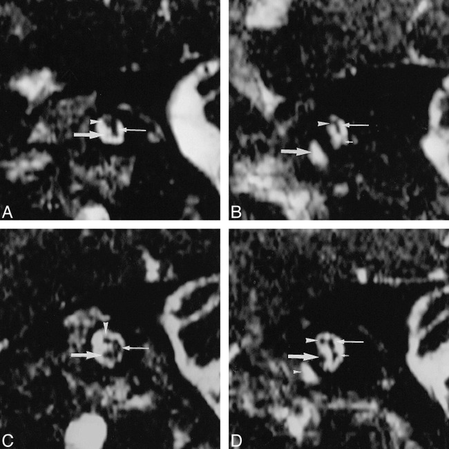
Images from the case of a 12-year-old female patient who presented at our institute with unilateral hearing impairment. A, Abnormal left ear. Parasagittal 3DFT-CISS (12.3/5.9/1; flip angle, 70°) reconstruction images obtained perpendicular to the IAC at the midportion in the affected left side. The facial nerve is depicted in the superior and anterior position in the IAC (arrowhead). The common vestibular nerve is identified (thin arrow). A thin cochlear branch arises (arrow) from the VCN. B, Abnormal left ear. Parasagittal 3DFT-CISS reconstruction images obtained perpendicular to the IAC at the lateral portion. The common vestibular nerve (not identified on this image) divides into the superior vestibular branch (thin arrow) and the inferior vestibular branch (short arrow) (poorly visualized). The cochlear branch is not visualized. The IAC seems to be slightly narrow compared with that on the right side (see panel D). The facial nerve is identified (arrowhead). The basal turn of the cochlea is shown (arrow). The vestibular branches also seem to be small in comparison with those on the right side (see panel D). Any vestibular abnormality, however, could be detected in the patient. C, Normal right ear. Parasagittal 3DFT-CISS (12.3/5.9/1; flip angle, 70°) reconstruction images obtained perpendicular to the IAC at the midportion in the normal right side. The cochlear branch (arrow) is larger than the facial nerve (arrowhead). The common vestibular nerve is shown (thin arrow). D, Normal right ear. Parasagittal 3DFT-CISS reconstruction images obtained perpendicular to the IAC at the lateral portion. The common vestibular nerve divides into the superior vestibular branch (thin arrow) and the inferior vestibular branch (short arrow). The cochlear branch (arrow) is larger than the facial nerve (arrowhead). Small arrowhead, the cochlea.
References
-
- Casselman JW, Kuhweide R. Ampe W, et al. Inner ear malformations in patients with sensorineural hearing loss: detection with gradient-echo (3DFT-CISS) MRI. Neuroradiology 1996;38:278-286 - PubMed
-
- Casselman JW, Offeciers FE, Govaerts PJ, et al. Aplasia and hypoplasia of the vestibulocochlear nerve: diagnosis with MR imaging. Radiology 1997;202:773-781 - PubMed
Publication types
MeSH terms
LinkOut - more resources
Full Text Sources
Medical