. 2000 Mar;66(3):895-903.
doi: 10.1128/AEM.66.3.895-903.2000.

Genetic analysis of chromosomal regions of Lactococcus lactis acquired by recombinant lytic phages

Affiliations

Genetic analysis of chromosomal regions of Lactococcus lactis acquired by recombinant lytic phages

E Durmaz et al. Appl Environ Microbiol. 2000 Mar.

Abstract

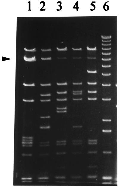
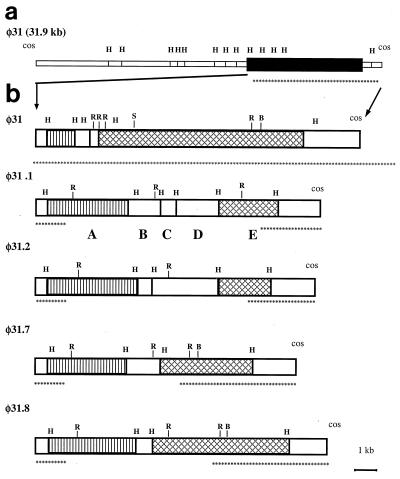
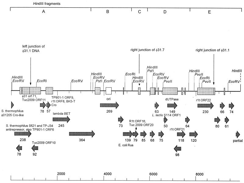
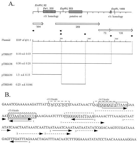
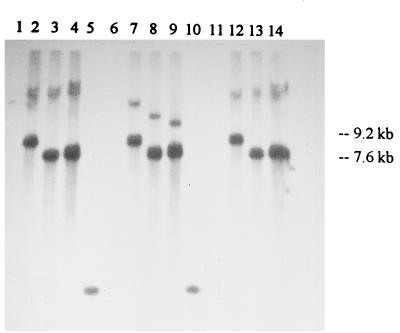
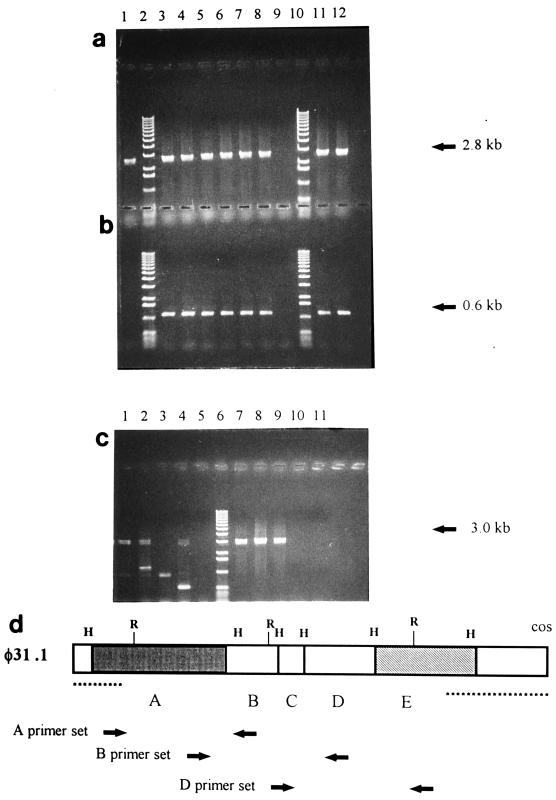
Recombinant phages are generated when Lactococcus lactis subsp. lactis harboring plasmids encoding the abortive type (Abi) of phage resistance mechanisms is infected with small isometric phages belonging to the P335 species. These phage variants are likely to be an important source of virulent new phages that appear in dairy fermentations. They are distinguished from their progenitors by resistance to Abi defenses and by altered genome organization, including regions of L. lactis chromosomal DNA. The objective of this study was to characterize four recombinant variants that arose from infection of L. lactis NCK203 (Abi(+)) with phage phi31. HindIII restriction maps of the variants (phi31.1, phi31.2, phi31.7, and phi31.8) were generated, and these maps revealed the regions containing recombinant DNA. The recombinant region of phage phi31.1, the variant that occurred most frequently, was sequenced and revealed 7.8 kb of new DNA compared with the parent phage, phi31. This region contained numerous instances of homology with various lactococcal temperate phages, as well as homologues of the lambda recombination protein BET and Escherichia coli Holliday junction resolvase Rus, factors which may contribute to efficient recombination processes. A sequence analysis and phenotypic tests revealed a new origin of replication in the phi31.1 DNA, which replaced the phi31 origin. Three separate HindIII fragments, accounting for most of the recombinant region of phi31.1, were separately cloned into gram-positive suicide vector pTRK333 and transformed into NCK203. Chromosomal insertions of each plasmid prevented the appearance of different combinations of recombinant phages. The chromosomal insertions did not affect an inducible prophage present in NCK203. Our results demonstrated that recombinant phages can acquire DNA cassettes from different regions of the chromosome in order to overcome Abi defenses. Disruption of these regions by insertion can alter the types and diversity of new phages that appear during phage-host interactions.


Figures

FIG. 1
FIG. 1
HindIII restriction digestion of φ31 (lane 1), φ31.1 (lane 2), φ31.2 (lane 3), φ31.7 (lane 4), and 31.8 (lane 5). Lane 6 contained a 1-kb marker (Gibco-BRL, Gaithersburg, Md.). The φ31 7-kb band (doublet) which is replaced in the recombinant phages is indicated by an arrowhead.
FIG. 2
FIG. 2
(a) HindIII map of the complete φ31 genome. The solid box represents the region replaced in the recombinant variants, which is expanded in panel b. (b) HindIII maps of φ31 and variants φ31.1, φ31.2, φ31.7, and φ31.8, showing the recombinant regions. Similar boxes indicate HindIII fragments that exhibit DNA homology, as determined by Southern blotting. HindIII fragments found in recombinant φ31.1 are labeled A through E. The sites marked H, R, B, and S are restriction sites for HindIII, EcoRI, BamHI, and SalI, respectively. The asterisks identify areas identical to areas in φ31, as determined by restriction mapping.
FIG. 3
FIG. 3
Genetic map of the recombinant region of φ31.1. The stipplied boxes indicate regions of DNA level homology with temperate lactococcal phages. The arrows indicate ORFs predicted from the φ31.1 sequence, and the numbers of amino acids are indicated below the arrows. Amino acid level homologies to other phage sequences are also indicated. The vertical arrows indicate junctions of the recombinant regions.
FIG. 4
FIG. 4
Localization of the putative origin region of phage φ31.1. (A) Schematic drawing of the region, showing predicted ORFs and regions of phage rlt homology. The small arrows indicate the positions of oligo primers used in PCR experiments to create subclones. The bars indicate the fragments cloned into pTRKH2. The EOPs are averages based on three or four replicates. (B) Sequence of the 240-bp shaded ori region. The solid arrows indicate indirect repeats, the dashed arrows indicate direct repeats, and the boxes indicate the stem regions of predicted stem-loops regions. s.d., standard deviation.
FIG. 5
FIG. 5
Southern hybridization showing insertion of the pTRK333-A, pTRK333-B, and pTRK333-C suicide plasmids into the chromosome of NCK203. Genomic DNA from NCK203 (lanes 1, 6, and 11), NCK203-A (lanes 2, 7, and 12), through NCK203-B (lanes 3, 8, and 13), or NCK203-D (lanes 4, 9, and 14) was digested with BamHI (lanes 1 through 4), PvuI (lanes 6 through 9), or SalI (lanes 11 through 14). Lanes 5 and 10 contained markers (one low-molecular-weight band in the marker lanes did hybridize with the probe). The Southern blot was probed with 32P-labeled pTRK333 DNA.
FIG. 6
FIG. 6
PCR products, showing that the pTRK333A chromosomal insertion did not occur in the region contributing to φ31.1. (a and b) Template DNA from NCK203 (lane 1), six single-colony isolates of NCK203-A (lanes 3 through 8), NCK203-B (lane 11), or NCK203-D (lane 12) and markers (lanes 2 and 10). Lane 9 contained a PCR control without template DNA. The primers used were set A (a) (expected product size, 2.8 kb) and the vector set (b) (expected product size, 0.6 kb). (c) Template DNA from NCK203 (lanes 1 and 7), NCK203-A (lanes 2 and 8), NCK203-B (lanes 3 and 9), or NCK203-D (lanes 4 and 10) and markers (lane 6). Lanes 5 and 11 contained a PCR control without template DNA. The primers used were set B (lanes 1 through 5) (expected product size, 3.0 kb) and set D (lanes 7 through 11) (expected product size, 3.1 kb). The smaller products obtained with primer set B were artifacts of one of the primers, which annealed to the vector, pTRK333. (d) Map of the recombinant region of φ31.1, similar to the map in Fig. 2b, showing the locations of the primer sets designed and used for the PCR experiment.

Similar articles

Cited by

References

    1. Alatossava T, Klaenhammer T R. Molecular characterization of three small isometric-headed bacteriophages which vary in their sensitivity to the lactococcal phage resistance plasmid pTR2030. Appl Environ Microbiol. 1991;57:1346–1353. - PMC - PubMed
    1. Birkeland N-K, Lonneborg A-M. The cos region of lactococcal bacteriophage φLC3. DNA Sequence J. 1993;4:211–214. - PubMed
    1. Boyce J D, Davidson B E, Hillier A J. Sequence analysis of the Lactococcus lactis temperate bacteriophage BK5-T and demonstration that the phage DNA has cohesive ends. Appl Environ Microbiol. 1995;61:4089–4098. - PMC - PubMed
    1. Chandry P S, Moore S C, Boyce J D, Davidson B E, Hillier A J. Analysis of the DNA sequence, gene expression, origin of replication and modular structure of the Lactococcus lactis lytic bacteriophage sk1. Mol Microbiol. 1997;26:49–64. - PubMed
    1. Dao M L, Ferretti J J. Streptococcus-Escherichia coli shuttle vector pSA3 and its use in the cloning of streptococcal genes. Appl Environ Microbiol. 1985;49:115–119. - PMC - PubMed

Publication types

LinkOut - more resources