Molecular analysis of the pmo (particulate methane monooxygenase) operons from two type II methanotrophs
- PMID: 10698759
- PMCID: PMC91930
- DOI: 10.1128/AEM.66.3.966-975.2000
Molecular analysis of the pmo (particulate methane monooxygenase) operons from two type II methanotrophs
Abstract
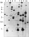
The particulate methane monooxygenase gene clusters, pmoCAB, from two representative type II methanotrophs of the alpha-Proteobacteria, Methylosinus trichosporium OB3b and Methylocystis sp. strain M, have been cloned and sequenced. Primer extension experiments revealed that the pmo cluster is probably transcribed from a single transcriptional start site located 300 bp upstream of the start of the first gene, pmoC, for Methylocystis sp. strain M. Immediately upstream of the putative start site, consensus sequences for sigma(70) promoters were identified, suggesting that these pmo genes are recognized by sigma(70) and negatively regulated under low-copper conditions. The pmo genes were cloned in several overlapping fragments, since parts of these genes appeared to be toxic to the Escherichia coli host. Methanotrophs contain two virtually identical copies of pmo genes, and it was necessary to use Southern blotting and probing with pmo gene fragments in order to differentiate between the two pmoCAB clusters in both methanotrophs. The complete DNA sequence of one copy of pmo genes from each organism is reported here. The gene sequences are 84% similar to each other and 75% similar to that of a type I methanotroph of the gamma-Proteobacteria, Methylococcus capsulatus Bath. The derived proteins PmoC and PmoA are predicted to be highly hydrophobic and consist mainly of transmembrane-spanning regions, whereas PmoB has only two putative transmembrane-spanning helices. Hybridization experiments showed that there are two copies of pmoC in both M. trichosporium OB3b and Methylocystis sp. strain M, and not three copies as found in M. capsulatus Bath.
Figures


Similar articles
-
Role of multiple gene copies in particulate methane monooxygenase activity in the methane-oxidizing bacterium Methylococcus capsulatus Bath.Microbiology (Reading). 1999 May;145 ( Pt 5):1235-1244. doi: 10.1099/13500872-145-5-1235. Microbiology (Reading). 1999. PMID: 10376840
-
Comparative analysis of the conventional and novel pmo (particulate methane monooxygenase) operons from methylocystis strain SC2.Appl Environ Microbiol. 2004 May;70(5):3055-63. doi: 10.1128/AEM.70.5.3055-3063.2004. Appl Environ Microbiol. 2004. PMID: 15128567 Free PMC article.
-
Particulate methane monooxygenase genes in methanotrophs.J Bacteriol. 1995 Jun;177(11):3071-9. doi: 10.1128/jb.177.11.3071-3079.1995. J Bacteriol. 1995. PMID: 7768803 Free PMC article.
-
Molecular genetics of methane oxidation.Biodegradation. 1994 Dec;5(3-4):145-59. doi: 10.1007/BF00696456. Biodegradation. 1994. PMID: 7765830 Review.
-
Molecular biology and regulation of methane monooxygenase.Arch Microbiol. 2000 May-Jun;173(5-6):325-32. doi: 10.1007/s002030000158. Arch Microbiol. 2000. PMID: 10896210 Review.
Cited by
-
Trace-gas metabolic versatility of the facultative methanotroph Methylocella silvestris.Nature. 2014 Jun 5;510(7503):148-51. doi: 10.1038/nature13192. Epub 2014 Apr 28. Nature. 2014. PMID: 24776799
-
Comparison of pmoA PCR primer sets as tools for investigating methanotroph diversity in three Danish soils.Appl Environ Microbiol. 2001 Sep;67(9):3802-9. doi: 10.1128/AEM.67.9.3802-3809.2001. Appl Environ Microbiol. 2001. PMID: 11525970 Free PMC article.
-
Boosting the acetol production in methanotrophic biocatalyst Methylomonas sp. DH-1 by the coupling activity of heteroexpressed novel protein PmoD with endogenous particulate methane monooxygenase.Biotechnol Biofuels Bioprod. 2022 Jan 17;15(1):7. doi: 10.1186/s13068-022-02105-1. Biotechnol Biofuels Bioprod. 2022. PMID: 35418298 Free PMC article.
-
The effect of lanthanum on growth and gene expression in a facultative methanotroph.Environ Microbiol. 2022 Feb;24(2):596-613. doi: 10.1111/1462-2920.15685. Epub 2021 Aug 12. Environ Microbiol. 2022. PMID: 34320271 Free PMC article.
-
Global Molecular Analyses of Methane Metabolism in Methanotrophic Alphaproteobacterium, Methylosinus trichosporium OB3b. Part II. Metabolomics and 13C-Labeling Study.Front Microbiol. 2013 Apr 3;4:70. doi: 10.3389/fmicb.2013.00070. eCollection 2013. Front Microbiol. 2013. PMID: 23565113 Free PMC article.
References
-
- Altschul S F, Gish W, Miller W, Myer E W. Basic local alignment search tool. J Mol Biol. 1990;215:403–410. - PubMed
-
- Bowman J P, Sly L I, Nichols P D, Hayward A C. Revised taxonomy of the methanotrophs: description of Methylobacter gen. nov., validation of Methylosinus and Methylocystis species, and a proposal that the family Methylococcaceae includes only the group I methanotrophs. Int J Syst Bacteriol. 1993;43:735–753.
-
- Brusseau G A, Tsien H C, Hanson R S, Wackett L P. Optimization of trichloroethylene oxidation by methanotrophs and use of a colorimetric assay to detect soluble methane monooxygenase activity. Biodegradation. 1990;1:19–29. - PubMed
-
- Dalton H, Wilkins P C, Jiang Y. Structure and mechanism of action of the hydroxylase of soluble methane monooxygenase. In: Murrell J C, Kelly D P, editors. Microbial growth on C1 compounds. Andover, United Kingdom: Intercept Ltd; 1993. pp. 65–80.
Publication types
MeSH terms
Substances
Associated data
- Actions
- Actions
LinkOut - more resources
Full Text Sources
Other Literature Sources

