. 2000 Mar;66(3):1038-49.
doi: 10.1128/AEM.66.3.1038-1049.2000.

Highly ordered vertical structure of Synechococcus populations within the one-millimeter-thick photic zone of a hot spring cyanobacterial mat

Collaborators, Affiliations

Highly ordered vertical structure of Synechococcus populations within the one-millimeter-thick photic zone of a hot spring cyanobacterial mat

N B Ramsing et al. Appl Environ Microbiol. 2000 Mar.

Abstract

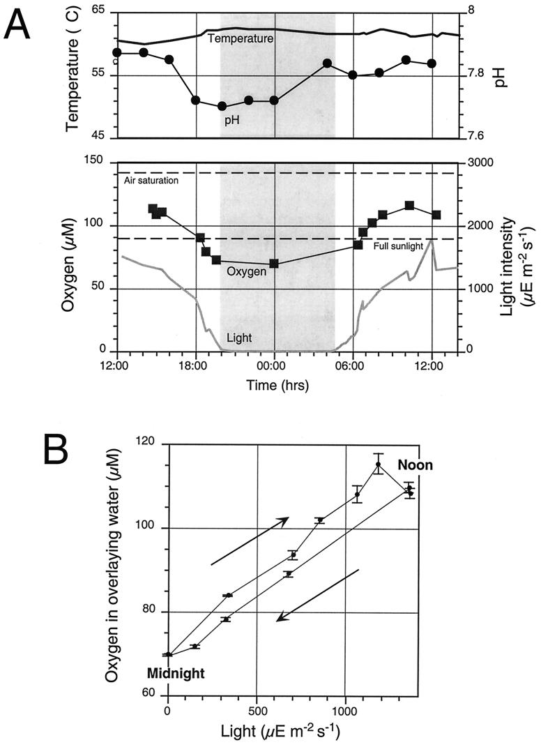
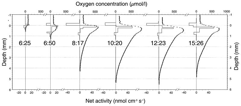
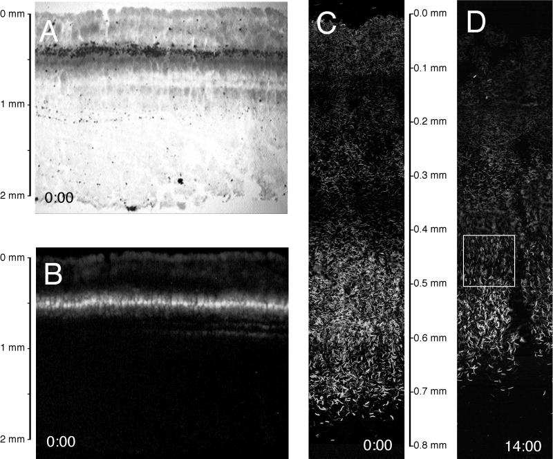
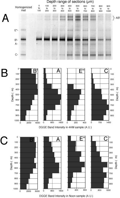
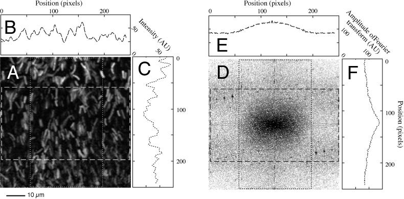
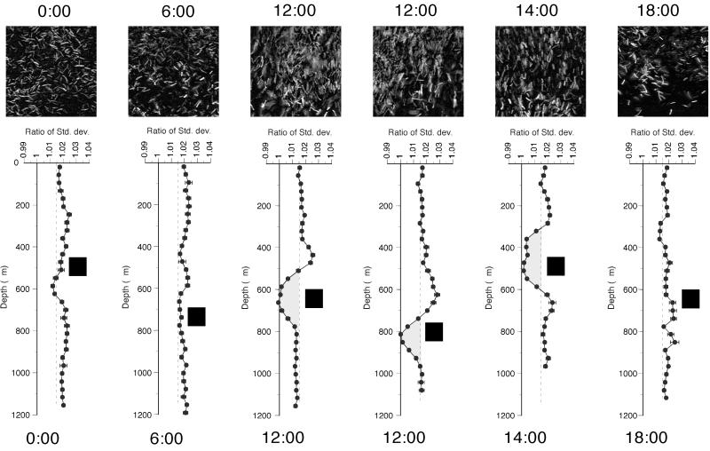
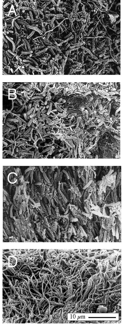
A variety of contemporary techniques were used to investigate the vertical distribution of thermophilic unicellular cyanobacteria, Synechococcus spp., and their activity within the upper 1-mm-thick photic zone of the mat community found in an alkaline siliceous hot spring in Yellowstone National Park in Wyoming. Detailed measurements were made over a diel cycle at a 61 degrees C site. Net oxygenic photosynthesis measured with oxygen microelectrodes was highest within the uppermost 100- to 200-microm-thick layer until midmorning, but as the day progressed, the peak of net activity shifted to deeper layers, stabilizing at a depth of 300 microm from midday throughout the afternoon. Examination of vertical thin sections by bright-field and autofluorescence microscopy revealed the existence of different populations of Synechococcus which form discrete bands at different vertical positions. Denaturing gradient gel electrophoresis analysis of PCR-amplified 16S rRNA gene segments from horizontal cryosections obtained at 100-microm-thick vertical intervals also suggested vertical stratification of cyanobacterial, green sulfur bacterium-like, and green nonsulfur bacterium-like populations. There was no evidence of diel migration. However, image analysis of vertical thin sections revealed the presence of a narrow band of rod-shaped Synechococcus cells in which the cells assumed an upright position. These upright cells, located 400 to 800 microm below the surface, were observed only in mat samples obtained around noon. In mat samples obtained at other time points, the cells were randomly oriented throughout the mat. These combined observations reveal the existence of a highly ordered structure within the very thin photic zone of this hot spring microbial mat, consisting of morphologically similar Synechococcus populations that are likely to be differentially adapted, some co-occurring with green sulfur bacterium-like populations, and all overlying green nonsulfur bacterium-like populations.


Figures

FIG. 1
FIG. 1
Diel variation in water chemistry above the 61°C cyanobacterial mat in Mushroom Spring Yellowstone National Park in Wyoming. (A) Changes in oxygen concentration, pH, temperature, and light intensity over the course of a day. The time span from sunset to sunrise is indicated by gray shading. (B) Correlation between oxygen concentration and light intensity, indicating lower net photosynthetic oxygen production in the afternoon. Reference time points are noted on the graph. Arrows indicate direction of progression during the diel cycle. Error bars show 95% confidence limits for the mean values reported. μE, microeinsteins.
FIG. 2
FIG. 2
Vertical stratification of metabolic processes within the Mushroom Spring 61°C Synechococcus mat from early morning to midafternoon. The small solid circles are the measurement points of in situ oxygen concentration profiles recorded at different times of the day with microelectrodes. The bars show the calculated net rates of oxygen production and consumption based on the observed concentration profiles and a simple diffusion model using a constant diffusion coefficient. Negative values indicate net consumption. The thin lines show the modeled concentration profiles according to the activity zonation indicated by the bars.
FIG. 3
FIG. 3
Images from 40-μm-thick vertical slices through a 61°C Mushroom Spring microbial mat sample at midnight (0:00) shown under bright-field (A) and epifluorescence (B) microscopy. (C) Enlarged view of epifluorescence image at midnight (0:00). (D) Enlarged view of epifluorescence image at 14:00. The boxed area in panel D is shown at a higher magnification in Fig. 5A.
FIG. 4
FIG. 4
Vertical distribution of 16S rRNA-defined genotypes in the 61°C Mushroom Spring microbial mat. (A) DGGE gel of the upper 2-mm cyanobacterial layer and of 100-μm-thick horizontal cryostat sections through the upper 1 mm of the mat's surface layer sampled at 04:00. Single letters correspond to Octopus Spring 16S rRNA sequence types: A and B′, cyanobacterial; C, green nonsulfur bacteria-like; E", green sulfur bacteria-like. AB′ bands are heteroduplex molecules (see text). (B and C) Bar graphs show the intensities of individual DGGE bands shown in panel A with depth from samples collected at night (04:00) (B) and at noon (12:00) (C). The intensities are depicted in arbitrary units (A.U.).
FIG. 5
FIG. 5
Estimating average cell orientation in the Mushroom Spring 61°C mat at 14:00 based on a two-dimensional fast Fourier transformation and subsequent analysis of the frequency distribution. (A) High magnification of part of a vertical transect through a 14:00 h sample (same as the boxed region of Fig. 3D). (B) Horizontal intensity profile through the part of panel A delineated by the horizontal rectangle. (C) Vertical intensity profile through the part of panel A delineated by the vertical rectangle. (D) Amplitude of Fourier transform of panel A. (E) Horizontal amplitude profile through horizontal rectangle in Fourier transform shown in panel D. (F) Vertical intensity profile through vertical rectangle in Fourier transform shown in panel D. For more extensive explanation, see Results.
FIG. 6
FIG. 6
Orientation of Synechococcus cells with depth in the Mushroom Spring 61°C mat expressed as the standard deviation of the Fourier transform in the vertical plane divided by the standard deviation of the Fourier Transform in the horizontal plane. Two different transects through a noon sample (12:00) show sample-to-sample variation. The micrographs above the analysis result are sample pictures taken at the depths indicated by black squares in the representative analysis profiles. The dashed lines represent the overall average ratio of 1.015. Values of <1.015, accentuated by the shaded area in the midday transects, denote the presence of vertically oriented cells.
FIG. 7
FIG. 7
Scanning electron microscopy picture of vertical cryosections of samples from the Mushroom Spring 61°C mat collected at noon. (A) Surface view with randomly oriented sausage-shaped Synechococcus cells. (B) Area near the surface (approximately 100 μm deep) also showing randomly oriented cells. (C) Vertically oriented Synechococcus cells found 700 μm below surface (D) Network of filamentous Chloroflexus-like organisms in undermat viewed from below. The scale bar in panel D applies to all panels.

References

    1. Bateson M M, Ward D M. Photoexcretion and fate of glycolate in a hot spring cyanobacterial mat. Appl Environ Microbiol. 1988;54:1738–1743. - PMC - PubMed
    1. Bauld J, Brock T D. Ecological studies of Chloroflexus, a gliding photosynthetic bacterium. Arch Microbiol. 1973;92:267–284.
    1. Bebout B M, Garcia-Pichel F. UV B-induced vertical migrations of cyanobacteria in a microbial mat. Appl Environ Microbiol. 1995;61:4215–4222. - PMC - PubMed
    1. Brock T D. Thermophilic microorganisms and life at high temperature. New York, N.Y: Springer Verlag; 1978.
    1. Castenholz R W. Ecology of blue-green algae in hot springs. In: Carr N G, Whitton B A, editors. The biology of blue-green algae. Vol. 1. Oxford, United Kingdom: Blackwell; 1973. pp. 379–414.

Publication types

LinkOut - more resources