

Abnormal angular gyrus asymmetry in schizophrenia
- PMID: 10698820
- PMCID: PMC2846293
- DOI: 10.1176/appi.ajp.157.3.428
Abnormal angular gyrus asymmetry in schizophrenia
Abstract
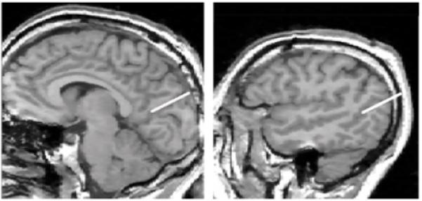
Objective: Few studies have evaluated the parietal lobe in schizophrenia despite the fact that it has an important role in attention, memory, and language-all functions that have been reported to be abnormal in schizophrenia. The inferior parietal lobule, in particular, is of interest because it is not only part of the heteromodal association cortex but also is part of the semantic-lexical network, which also includes the planum temporale. Both the inferior parietal lobule, particularly the angular gyrus of the inferior parietal lobule, and the planum temporale are brain regions that play a critical role as biological substrates of language and thought. The authors compared volume and asymmetry measures of the individual gyri of the parietal lobe by means of magnetic resonance imaging (MRI) scans.
Method: MRI scans with a 1. 5-Tesla magnet were obtained from 15 male chronic schizophrenic and 15 comparison subjects matched for age, gender, and parental socioeconomic status.
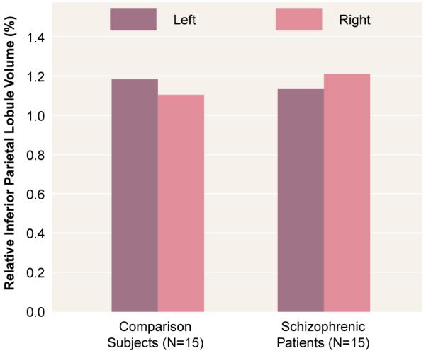
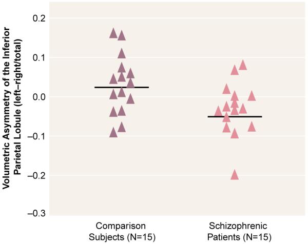
Results: Inferior parietal lobule volumes showed a leftward asymmetry (left 7.0% larger than right) in comparison subjects and a reversed asymmetry (left 6.3% smaller than right) in schizophrenic subjects. The angular gyrus accounted for this difference in asymmetry, with the left angular gyrus being significantly larger (18.7%) than the right in comparison subjects, a finding that was not observed in schizophrenic patients. A further test of angular gyrus asymmetry showed a reversal of the normal left-greater-than-right asymmetry in the schizophrenic patients.
Conclusions: Patients with schizophrenia showed a reversed asymmetry in the inferior parietal lobule that was localized to the angular gyrus, a structure belonging to the heteromodal association cortex as well as being part of the semantic-lexical network. This finding contributes to a more comprehensive understanding of the neural substrates of language and thought disorder in schizophrenia.
Figures
References
-
- Mesulam MM. Large-scale neurocognitive networks and distributed processing for attention, language, and memory. Ann Neurol. 1990;28:597–613. - PubMed
-
- Park S, Holzman PS. Schizophrenics show spatial working memory deficits. Arch Gen Psychiatry. 1992;49:975–982. - PubMed
-
- Nuechterlein KH, Dawson ME. Information processing and attentional functioning in the developmental course of schizophrenic disorders. Schizophr Bull. 1984;10:160–203. - PubMed
-
- Nestor PG, Faux SF, McCarley RW, Shenton ME, Sands SF. Measurement of visual sustained attention in schizophrenia using signal detection analysis and a newly developed computerized CPT task. Schizophr Res. 1990;3:329–332. - PubMed
-
- Shenton ME, Wible CG, McCarley RW. A review of magnetic resonance imaging studies of brain abnormalities in schizophrenia. In: Krishnan KRR, Doraiswamy PM, editors. Brain Imaging in Clinical Psychiatry. Marcel Dekker; New York: 1997. pp. 297–380.
Publication types
MeSH terms
Grants and funding
LinkOut - more resources
Full Text Sources
Medical
