. 2000 Mar 1;19(5):807-18.
doi: 10.1093/emboj/19.5.807.

Crystal structure of ribosomal protein L4 shows RNA-binding sites for ribosome incorporation and feedback control of the S10 operon

Affiliations

Crystal structure of ribosomal protein L4 shows RNA-binding sites for ribosome incorporation and feedback control of the S10 operon

M Worbs et al. EMBO J. .

Abstract

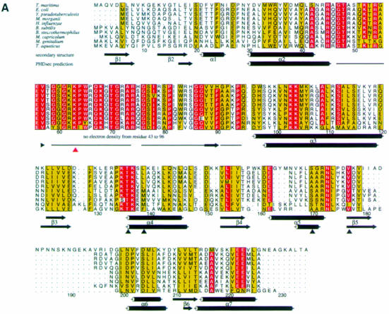
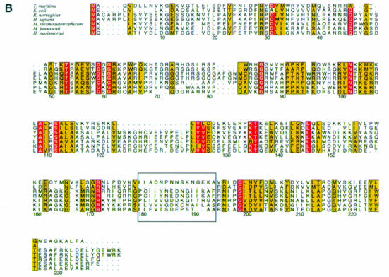
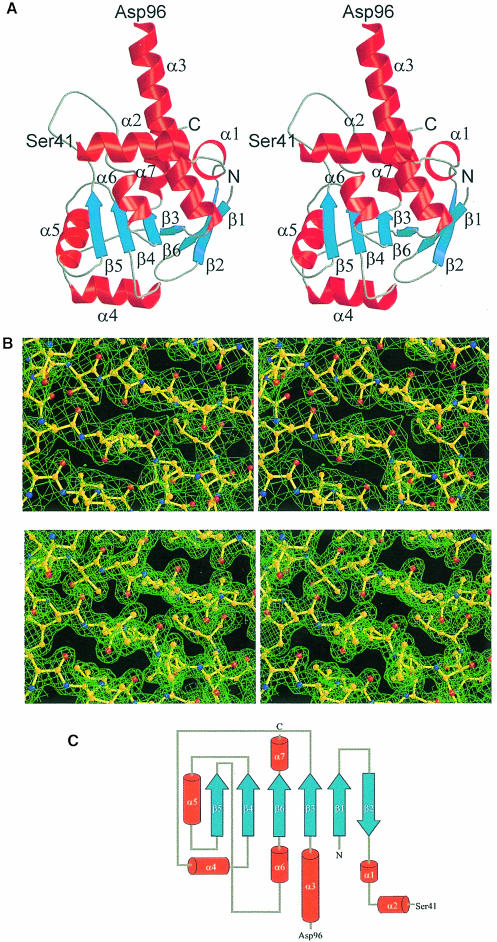
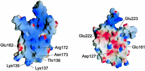
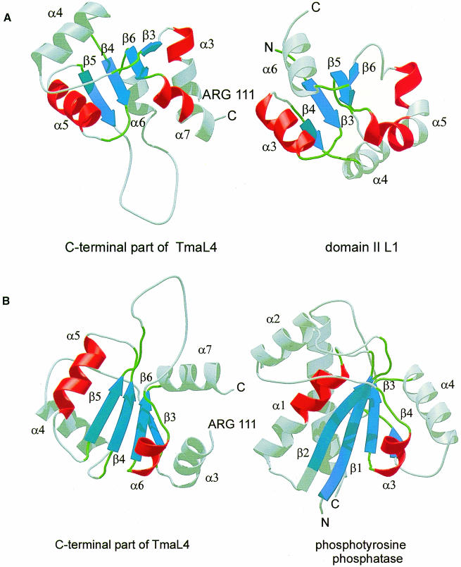
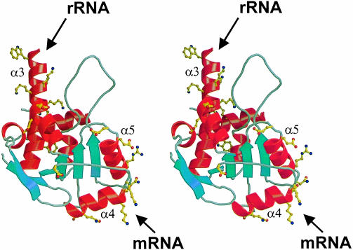
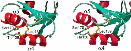
Ribosomal protein L4 resides near the peptidyl transferase center of the bacterial ribosome and may, together with rRNA and proteins L2 and L3, actively participate in the catalysis of peptide bond formation. Escherichia coli L4 is also an autogenous feedback regulator of transcription and translation of the 11 gene S10 operon. The crystal structure of L4 from Thermotoga maritima at 1.7 A resolution shows the protein with an alternating alpha/beta fold and a large disordered loop region. Two separate binding sites for RNA are discernible. The N-terminal site, responsible for binding to rRNA, consists of the disordered loop with flanking alpha-helices. The C-terminal site, a prime candidate for the interaction with the leader sequence of the S10 mRNA, involves two non-consecutive alpha-helices. The structure also suggests a C-terminal protein-binding interface, through which L4 could be interacting with protein components of the transcriptional and/or translational machineries.


Figures

None
Fig. 1. (A) Alignment of representative L4 sequences from different bacteria. The species are as follows: Thermotoga maritima (Nelson et al., 1999); Escherichia coli (Zurawski and Zurawski, 1985); Yersinia pseudotuberculosis (Gross et al., 1989); Morganella morganii (Zengel et al., 1995); Haemophilus influenzae (Fleischmann et al., 1995); Bacillus subtilis (Yasumoto et al., 1996); Bacillus stearothermophilus (Herwig et al., 1992); Mycoplasma capricolum (Ohkubo et al., 1987); Mycoplasma genitalium (Fraser et al., 1995); and Thermus aquaticus (Pfeiffer et al., 1995). The alignment was performed using PILEUP [Wisconsin Package Version 9.0, Genetics Computer Group (GCG), Madison, WI] and drawn with the program ALSCRIPT (Barton, 1993). The numbering corresponds to TmaL4. The background of amino acids strictly conserved in at least nine out of 10 species is colored red. Residues with conservation values >5 in at least nine sequences are drawn with a yellow background (Livingstone and Barton, 1993). The secondary structures as determined by the program STRIDE (Frishman and Argos, 1995) and the corresponding PHDsec secondary structure predictions (Rost and Sander, 1993) are also given. Predicted coil regions (horizontal line) are shown just from amino acids 43–96. Residues evaluated as important for the regulatory functions of TmaL4 (Li et al., 1996) are indicated by black triangles, whereas the site conferring erythromycin resistance in the case of mutation (Chittum and Champney, 1994) is indicated with a red triangle. (B) Sequence alignment of L4 proteins from the three kingdoms of life. The following species were used: Thermotoga maritima (Nelson et al., 1999); Escherichia coli (Zurawski and Zurawski, 1985); Rattus norvegicus (Chan et al., 1995); Homo sapiens (Bagni et al., 1993); Methanobacterium thermoautotrophicum (Smith et al., 1997); Methanococcus jannaschii (Bult et al., 1996); and Haloarcula marismortui (Arndt et al., 1990). The area of the extra loop of TmaL4 and the corresponding sequences from the other proteins is boxed. The color coding is the same as in (A). The C–terminal extension of ∼180 amino acids, which is typical for eukaryotic L4 proteins, is omitted from the alignment.
None
Fig. 2. (A) Stereo ribbon diagram of TmaL4 showing the overall fold. The secondary structural elements are labeled according to Figure 1A. Unless indicated otherwise, figures were produced with MOLSCRIPT (Kraulis, 1991) and rendered with Raster3D (Merritt and Bacon, 1997). (B) A stereo view of a portion of the electron density around the four-stranded central β-sheet in the C–terminus of the protein. The top part shows the solvent-flattened MIRAS map calculated at 2.5 Å contoured at 0.8σ. The bottom part displays the final 2FoFc map at 1.7 Å contoured at 1.4σ. (C) Topology diagram of TmaL4. Color coding is the same as in (A).
None
Fig. 3. Surface electrostatic potential of TmaL4. The figure on the left shows the putative RNA-binding surface. The positions of important and conserved surface residues are indicated. The figure on the right corresponds to the probable protein-binding site on the opposite side of the molecule. The potentials were calculated with the program GRASP (Nicholls et al., 1991).
None
Fig. 4. (A) Comparison of the C–terminus of TmaL4 (left) with domain II of r–protein L1 (right; Nikonov et al., 1996). Corresponding parts are in the same color. (B) Ribbon diagrams showing the C–terminus of TmaL4 and phosphotyrosine protein phosphatase (Su et al., 1994). Secondary structural elements that could be aligned again are in the same color.
None
Fig. 5. Stereo view of TmaL4. Conserved residues are shown in ball-and-stick representation. The spatial separation of the different functional sites is clearly seen. The long helix α3 (top) and helix α2 (behind α3, in the background) harbor some of the amino acids implicated in interactions with rRNA. The probable mRNA-binding part of the molecule in the C–terminus is located in helices α4 and α5 (bottom, on the right side of the molecule). In the foreground, some conserved residues belonging to the putative protein-binding site are seen.
None
Fig. 6. Stereo ribbon plot of the regulative part of the molecule encompassing helices α4 and α5. The four amino acids known to be essential for regulation (Li et al., 1996) are drawn in ball-and-stick representation. Leu139 and Val177 are pointing inwards to the hydrophobic core of TmaL4. The side chain of Ser170 stabilizes helix α5, and Thr136 (in the foreground) protrudes from the molecule.

Similar articles

Cited by

References

    1. Agafonov D.E., Kolb, V.A. and Spirin, A.S. (1997) Proteins on ribosome surface: measurements of protein exposure by hot tritium bombardment technique. Proc. Natl Acad. Sci. USA, 94, 12892–12897. - PMC - PubMed
    1. Albright R.A. and Matthews, B.W. (1998) Crystal structure of λ-Cro bound to a consensus operator at 3.0 Å resolution. J. Mol. Biol., 280, 137–151. - PubMed
    1. Allen T., Shen, P., Samsel, L., Liu, R., Lindahl, L. and Zengel, J.M. (1999) Phylogenetic analysis of L4-mediated autogenous control of the S10 ribosomal protein operon. J. Bacteriol., 181, 6124–6132. - PMC - PubMed
    1. Arndt E., Kromer, W. and Hatakeyama, T. (1990) Organization and nucleotide sequence of a gene cluster coding for eight ribosomal proteins in the archaebacterium Halobacterium marismortui. J. Biol. Chem., 265, 3034–3039. - PubMed
    1. Bagni C., Mariottini, P., Annesi, F. and Amaldi, F. (1993) Human ribosomal protein L4: cloning and sequencing of the cDNA and primary structure of the protein. Biochim. Biophys. Acta, 1216, 475–478. - PubMed

Publication types

LinkOut - more resources