. 2000 Mar 1;19(5):819-30.
doi: 10.1093/emboj/19.5.819.

Crystal structure of the catalytic portion of human HMG-CoA reductase: insights into regulation of activity and catalysis

Affiliations

Crystal structure of the catalytic portion of human HMG-CoA reductase: insights into regulation of activity and catalysis

E S Istvan et al. EMBO J. .

Abstract

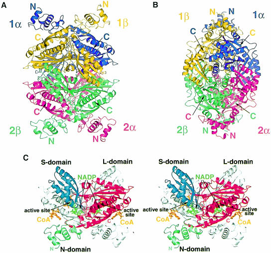
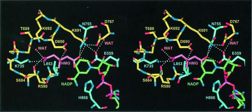
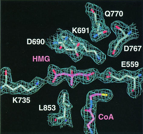
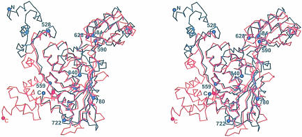
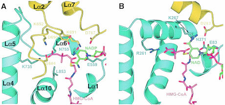
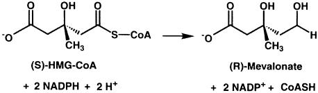
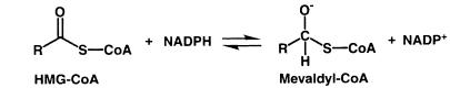
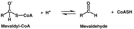
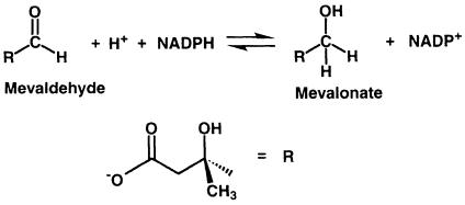
3-hydroxy-3-methylglutaryl-CoA reductase (HMGR) catalyzes the formation of mevalonate, the committed step in the biosynthesis of sterols and isoprenoids. The activity of HMGR is controlled through synthesis, degradation and phosphorylation to maintain the concentration of mevalonate-derived products. In addition to the physiological regulation of HMGR, the human enzyme has been targeted successfully by drugs in the clinical treatment of high serum cholesterol levels. Three crystal structures of the catalytic portion of human HMGR in complexes with HMG-CoA, with HMG and CoA, and with HMG, CoA and NADP(+), provide a detailed view of the enzyme active site. Catalytic portions of human HMGR form tight tetramers. The crystal structure explains the influence of the enzyme's oligomeric state on the activity and suggests a mechanism for cholesterol sensing. The active site architecture of human HMGR is different from that of bacterial HMGR; this may explain why binding of HMGR inhibitors to bacterial HMGRs has not been reported.


Figures

None
Fig. 1. Ribbon diagrams of human HMGR. (A and B) The structure of the HMGR homotetramer. Dimer 1 consists of the monomers called ‘1α’ (purple) and ‘1β’ (yellow), while dimer 2 consists of monomers ‘2α’ (pink) and ‘2β’ (green). (C) Stereo diagram of the HMGR dimer structure. For clarity, only one monomer is colored. The N–terminal N–domain is in green, the large L–domain is in red and the small S–domain is in blue. The N–terminus (residue 462) and the C–terminus (residue 870) for the colored monomer are indicated. CoA (orange) and NADP(H) (green) are labeled and the two active sites are marked. This figure and Figures 5–8 were prepared with Bobscript (Esnouf, 1999), gl_render (L.Esser, personal communication) and POV-Ray (Persistence of Vision Ray Tracer v3.02, Copyright 1997 POV-Team, http://www.povray.org).
None
Fig. 2. Topology diagram of the human HMGR monomer. Colors for the three domains are as in Figure 1C. Helices are shown as outlined rectangles and strands are shown as solid arrows. The central helix Lα10 in the L–domain is indicated by the outlined circle. Residues that participate in substrate binding as well as the N- and C–termini are indicated.
None
Fig. 3. Structure-based sequence alignment of human and P.mevalonii HMGR (PDB code 1qax; Tabernero et al., 1999). Colors of the secondary structure elements are as in Figure 1C; solid arrows indicate β–strands, outlined rectangles indicate α–helices and an asterisk indicates a 310-helix. Regions of high structural similarity are in yellow, residues involved directly in substrate binding are in purple and regions involved in dimerization or tetramerization are marked by the words ‘dimer’ and ‘tetramer’. The phosphorylation site (S872) of human HMGR is marked in green.
None
Fig. 4. Molecular surface of the dimer–dimer interface. (A) Atoms between 0.1 and 6 Å of the dimer–dimer interface are indicated by a color gradient of yellow to orange. (B) Colored according to residue type with hydrophobic residues (A, V, F, P, M, I, L, Y, W and G) in green, polar residues (S, T, H, C, N and Q) in white, K and R in blue, and D and E in red. This figure was prepared with GRASP (Nicholls et al., 1991), gl_render and POV-Ray.
None
Fig. 5. Stereo diagram showing residues involved in HMG binding based on the form C structure at a resolution of 2.0 Å. Residues from monomer α are in yellow and residues from monomer β are in blue. HMG is magenta and NADP+ is in green. All distances within 3.0 Å or closer are indicated by dotted lines.
None
Fig. 6. Simulated annealing omit 2FoFc map at 2.1 Å and contoured at 1σ showing the cleaved HMG and CoA of the form A structure. HMG and CoA are colored in magenta.
None
Fig. 7. Stereo drawing of the Cα superposition of human (in blue) and P.mevalonii (in red) (PDB code 1qax) HMGR monomer. The superposition was done with the program LSQMAN (Kleywegt, 1997). Selected residues in human HMGR are indicated by blue spheres and numbered. The N- and C–termini are indicated.
None
Fig. 8. Ribbon diagram of the human HMGR (A) and P.mevalonii HMGR (B) active site. Residues from monomer α are in yellow, residues from monomer β are in blue, HMG-CoA is in magenta and NAD(P) is in green. For human HMGR, secondary structure elements are labeled. For clarity, not all CoA atoms are shown. Side chains of selected residues are indicated.
None
Scheme 1.
None
Scheme 1.
None
Scheme 2.
None
Scheme 2.
None
Scheme 3.
None
Scheme 3.
None
Scheme 4.

References

    1. Beach M.J. and Rodwell, V.W. (1989) Cloning, sequencing and overexpression of mvaA, which encodes Pseudomonas mevalonii 3-hydroxy-3-methylglutaryl coenzyme A reductase. J. Bacteriol., 171, 2994–3001. - PMC - PubMed
    1. Bennis F., Favre, G., Le Gaillard, F. and Soula, G. (1993) Importance of mevalonate-derived products in the control of HMG-CoA reductase activity and growth of human lung adenocarcinoma cell line A549. Int. J. Cancer, 55, 640–645. - PubMed
    1. Blackwell J.R. and Horgan, R. (1991) A novel strategy for production of a highly expressed recombinant protein in an active form. FEBS Lett., 295, 10–12. - PubMed
    1. Bochar D.A., Stauffacher, C.V. and Rodwell, V.W. (1999a) Sequence comparisons reveal two classes of 3-hydroxy-3-methylglutaryl coenzyme A reductase. Mol. Genet. Metab., 66, 122–127. - PubMed
    1. Bochar D.A., Tabernero, L., Stauffacher, C.V. and Rodwell, V.W. (1999b) Aminoethylcysteine can replace the function of the essential active site lysine of Pseudomonas mevalonii 3-hydroxy-3-methylglutaryl coenzyme A reductase. Biochemistry, 38, 8879–8883. - PubMed

Substances