. 2000 Mar 1;19(5):862-9.
doi: 10.1093/emboj/19.5.862.

TRAPP stably associates with the Golgi and is required for vesicle docking

Affiliations

TRAPP stably associates with the Golgi and is required for vesicle docking

J Barrowman et al. EMBO J. .

Abstract

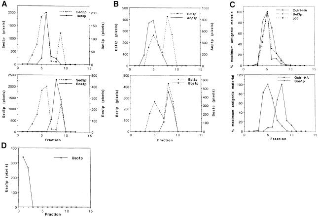
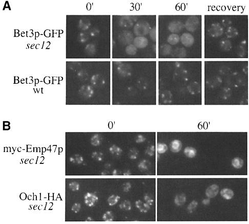
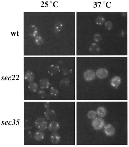
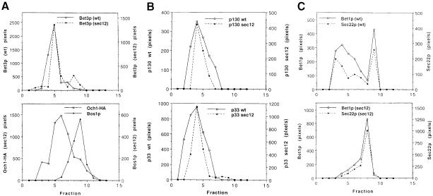
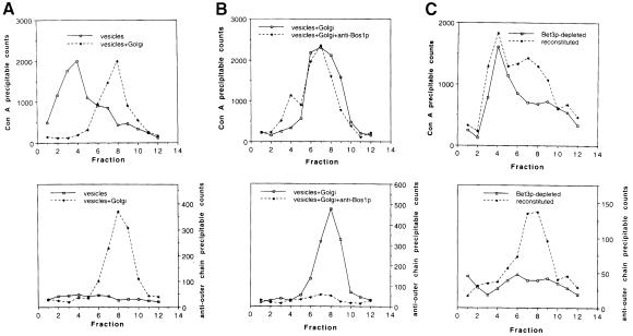
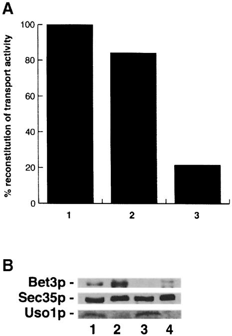
Bet3p, a component of a large novel complex called TRAPP, acts upstream of endoplasmic reticulum (ER)-Golgi SNAREs. Unlike the SNAREs, which reside on multiple compartments, Bet3p is localized exclusively to Golgi membranes. While other proteins recycle from the Golgi to the ER, Bet3p and other TRAPP subunits remain associated with this membrane under conditions that block anterograde traffic. We propose that the persistent localization of TRAPP to the Golgi may be important for its role in docking vesicles to this membrane. Consistent with this proposal, we find that transport vesicles fail to bind to Golgi membranes in vitro in the absence of Bet3p. Binding is restored by the addition of cytosol containing Bet3p. These findings indicate that TRAPP stably associates with the Golgi and is required for vesicle docking.


Figures

None
Fig. 1. Steady-state localization of TRAPP subunits, SNAREs and Uso1p on sucrose velocity gradients. Data in (C) and (D) are from separate gradients. (A) Top panel: Bet3p (solid line) co-fractionates with the Golgi peak of Sed5p (broken line). Bottom panel: the second peak of Sed5p (broken line) co-fractionates with Bos1p (solid line), an ER marker. (B) Top panel: the first peak of Bet1p (broken line) co-fractionates with Anp1p (solid line), an early Golgi marker. Bottom panel: the second peak of Bet1p (broken line) co-fractionates with Bos1p (solid line). (C) Top panel: TRAPP subunits Bet3p (♦) and p33 (▪) co-fractionate with Och1-HA (□), another early Golgi marker. Bottom panel: the peaks of Och1p (□) and Bos1p (♦) are distinct on this gradient, showing that the Golgi and ER are well resolved. (D) In contrast to TRAPP subunits, Uso1p (□) is largely soluble and is not detected on membranes.
None
Fig. 2. Bet3p–GFP and Och1-HA become finely punctate in sec12 at 37°C. Wild-type and sec12 cells, containing Bet3p–GFP, Och1-HA or myc-Emp47p, were shifted to 37°C for the indicated times followed by appropriate preparation for imaging under a Zeiss fluorescence light microscope. (A) Bet3p–GFP was imaged in live cells at the indicated time points ranging from 0 (cells at 25°C prior to shift) to 60 min. Following the 1 h incubation at 37°C, cells subsequently were shifted to 25°C and incubated in the presence of 20 μg/ml cycloheximide for 1 h to allow for recovery. The punctate signal from Bet3p–GFP becomes hazy after 30 min at 37°C, and returns upon a shift back to permissive temperature. (B) As controls, the localization of myc-Emp47p, a recycling Golgi protein, and Och1-HA was followed in sec12. The tagged proteins were detected using indirect immuno- fluorescence as described in Materials and methods. Och1-HA is detected on elongated punctate structures at the 0 min time point, which disperse after 60 min at 37°C. In contrast and as previously reported (Schröder et al., 1995), myc-Emp47p recycles to the ER under these conditions.
None
Fig. 3. Bet3p–GFP disperses in a fusion and tethering mutant. Wild-type, sec22 and sec35 strains containing Bet3p–GFP were shifted to 37°C for 1 h.
None
Fig. 4. Fractionation of Bet3p in wild-type and sec12 at 37°C. Wild-type and sec12 cells were shifted to 37°C for 1 h, followed by lysis and loading on sucrose gradients. Data in (A), (B) and (C) are from separate gradients. (A) Top panel: Bet3p fractionates similarly in wild-type (solid line) and sec12 (broken line) at 37°C. We consider the minor peak of Bet3p in fraction 8 to be insignificant as it was not reproduced in other gradients. Bottom panel: the peaks of Och1p (□) and Bos1p (♦) in lysates from sec12 are distinct, showing that Golgi and ER membranes are resolved in the mutant. (B) TRAPP subunits p130 (top panel) and p33 (bottom panel) also fractionate similarly in wild-type (solid line) and sec12 (broken line). (C) Top panel: Bet1p (solid line) and Sec22p (broken line) co-fractionate in Golgi and ER peaks in lysates from wild-type. Bottom panel: in lysates from sec12, Bet1p (solid line) and Sec22p (broken line) redistribute to the ER in sec12 at 37°C and are no longer detected in Golgi fractions.
None
Fig. 5. Fractions depleted of Bet3p in vivo fail to support the binding of ER-derived vesicles to Golgi membranes in vitro. Bet3p-depleted fractions were prepared and assayed as described previously (Groesch et al., 1992; Sacher et al., 1998). The reaction product was analyzed on a sucrose velocity gradient as described in Materials and methods. The slight changes made in the velocity gradient are sufficient to allow separation of ER–Golgi transport vesicles from Golgi membranes. (A) Top panel: free vesicles (solid line) and vesicles that have fused with the Golgi (broken line) migrate to distinct locations. Bottom panel: pro-α-factor is ubject to outer chain modification when vesicles are incubated in the presence of Golgi membranes (broken line), indicating that fusion with the Golgi has occurred. (B) Top panel: in the presence of anti-Bos1p antibodies (broken line), vesicles bind to Golgi membranes. Bottom panel: in the presence of anti-Bos1p antibodies, the vesicles that migrate with the Golgi have not fused (broken line), as indicated by the lack of outer chain modification. (C) Top panel: the depletion of Bet3p prevents vesicles from binding to Golgi membranes (solid line). The addition of cytosol containing Bet3p partially reconstitutes vesicle docking (broken line). Bottom panel: vesicle fusion is reconstituted by the addition of cytosol containing Bet3p, as indicated by the presence of outer chain modifications (broken line).
None
Fig. 6. Bet3p is required for the reconstitution of transport activity in vitro. (A) Reconstitution of ER to Golgi transport in vitro was measured with anti-outer chain antiserum. The data shown are representative. Untreated (column 1) or mock-treated cytosol (column 2) prepared from a strain (SFNY 904) containing Bet3p fused to protein A restored transport activity in vitro to the Bet3p-depleted Golgi. Treatment of cytosol from the tagged strain (SFNY904) with IgG–Sepharose reduced its ability to reconstitute activity (column 3). The activity of Bet3p-depleted cytosol assayed in the presence of Bet3p-depleted membranes was 15% that of wild-type fractions. A 1.7-fold increase in activity was observed in the reconstituted sample. (B) Wild-type and depleted fractions were blotted for the presence of Bet3p, Sec35p and Uso1p. Shown here are cytosolic (lanes 1 and 3) and membrane (lanes 2 and 4) fractions prepared from wild-type (lanes 1 and 2) and the Bet3p-depleted strain (lanes 3 and 4). The ability to detect Bet3p is significantly reduced in fractions from the Bet3p-depleted strain (compare lanes 1 and 2 with lanes 3 and 4), while the levels of Sec35p and Uso1p are unaffected. Lanes 1–4 contain equal amounts of protein (250 μg).

Similar articles

Cited by

References

    1. Antebi A. and Fink, R.G. (1992) The yeast Ca2+-ATPase homologue, PMR1, is required for normal Golgi function and localizes in a Golgi-like distribution. Mol. Biol. Cell, 3, 633–654. - PMC - PubMed
    1. Barlowe C. (1997) Coupled ER to Golgi transport reconstituted with purified cytosolic proteins. J. Cell Biol., 139, 1097–1108. - PMC - PubMed
    1. Barlowe C. and Schekman, R. (1993) SEC12 encodes a guanine-nucleotide-exchange factor essential for transport vesicle budding from the ER. Nature, 365, 347–349. - PubMed
    1. Berkower C., Loayza, D. and Michaelis, S. (1994) Metabolic instability and constitutive endocytosis of STE6, the α-factor transporter of Saccharomyces cerevisiae. Mol. Biol. Cell, 5, 1185–1198. - PMC - PubMed
    1. Bonangelino C.J., Catlett, N.L. and Weisman, L.S. (1997) Vac7p, a novel vacuolar protein, is required for normal vacuole inheritance and morphology. Mol. Biol. Cell, 17, 6847–6858. - PMC - PubMed

Publication types

LinkOut - more resources