. 2000 Mar 1;19(5):870-81.
doi: 10.1093/emboj/19.5.870.

The luminal part of the murine cytomegalovirus glycoprotein gp40 catalyzes the retention of MHC class I molecules

Affiliations

The luminal part of the murine cytomegalovirus glycoprotein gp40 catalyzes the retention of MHC class I molecules

H Ziegler et al. EMBO J. .

Abstract

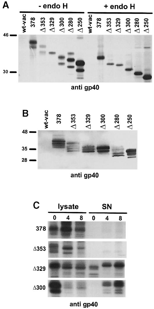
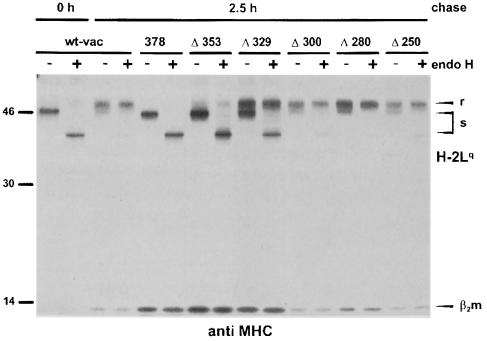
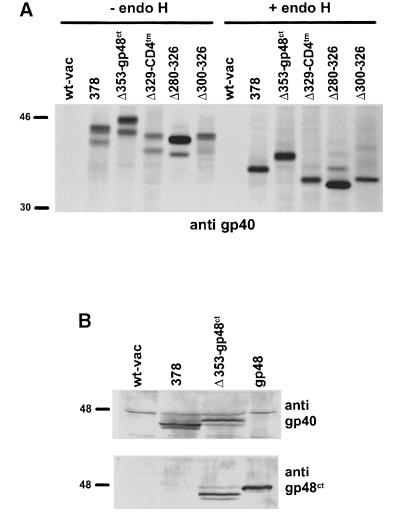
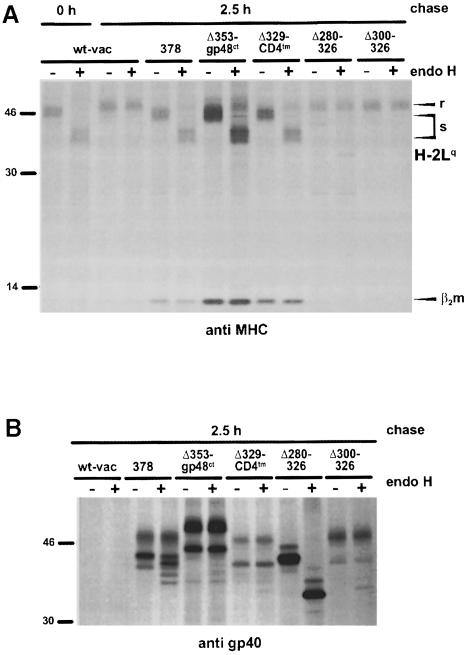
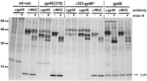
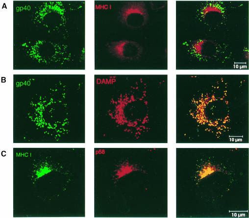
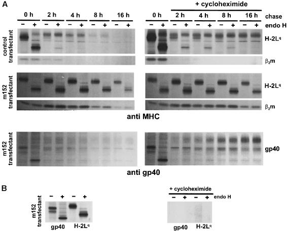
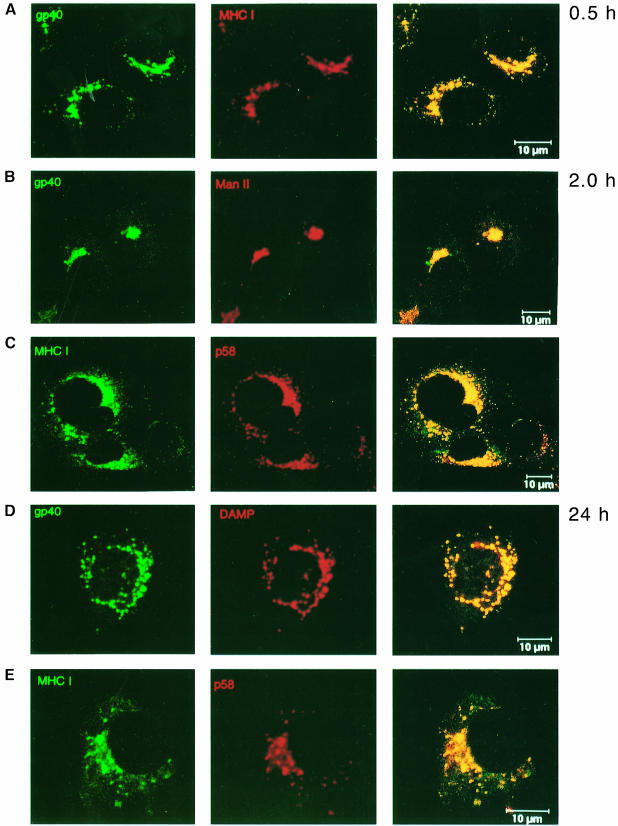
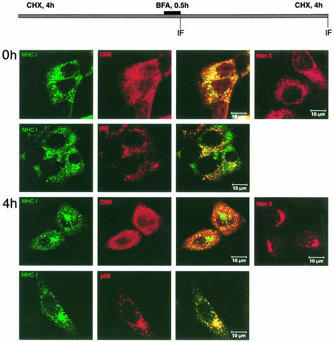
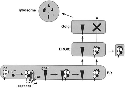
Murine cytomegalovirus (MCMV) interferes with the MHC class I pathway of antigen presentation. The type I transmembrane glycoprotein gp40, encoded by the gene m152, retains major histocompatibility complex (MHC) class I complexes in the endoplasmic reticulum (ER)-Golgi intermediate compartment (ERGIC)/cis-Golgi. These MHC class I complexes are stable, show an extended half-life and do not exchange beta(2)-microglobulin, whereas gp40 reaches an endosomal/lysosomal compartment where it subsequently is degraded. The analysis of regions within the viral protein that are essential for MHC class I retention revealed that a secreted form of gp40, lacking the cytoplasmic tail and the transmembrane region, still has the capacity to retain MHC class I complexes. Continuous expression of gp40 is not required for MHC class I retention. Our data indicate that the retention of MHC class I complexes in the ERGIC/cis-Golgi is triggered by gp40 and does not require the further presence of the viral protein.


Figures

None
Fig. 1. Schematic representation of gp40 deletion mutants and chimeric proteins. (A) Proportional representation of the m152 gene product gp40 (378) with signal peptide (sp), glycosylation sites (branched symbols), transmembrane region (tm) and cytoplasmic tail (ct). The bar indicates the binding region of the peptide antiserum to gp40 (p11). The number of the deletion mutants refers to the last amino acid expressed in the truncated protein. (B) The number in the chimeric proteins indicates the last amino acid of gp40 to which the cytoplasmic tail of gp48 (-gp48ct) or the transmembrane region of CD4 (-CD4tm) was fused. The bar indicates the binding region of the mAb to the cytoplasmic tail of gp48 (CROMA229). The numbers of the two intramolecular deletion mutants indicate the depleted region. (C) Properties of the mutant gp40 proteins described in this study: the release of the proteins into the culture supernatant, their transport to lysosomes and ability to block the transport of MHC class I molecules. nd, not determined.
None
Fig. 2. Expression of gp40 deletion mutants by recombinant vaccinia virus. (A) NIH 3T3 cells were infected with wild-type vaccinia virus (wt-vac), recombinant vaccinia virus expressing the complete m152 ORF (378) and the C-terminal deletion mutants (Δ353, Δ329, Δ300, Δ280 and Δ250). At 5 h post-infection, cells were pulse-labeled for 30 min with [35S]cysteine/methionine. The gp40 derivatives were precipitated with peptide antiserum to gp40 (p11). Half of the precipitates were either digested with endoglycosidase H (endo H) or mock treated before separation by 10% SDS–PAGE. (B) Western blot analysis of NIH 3T3 cells infected with wt-vac and the indicated recombinant vaccinia viruses, respectively. At 12 h post-infection, total cellular lysates were prepared and separated by 10% SDS–PAGE. After blotting, binding of the mAb against gp40 (gpM3D10) was visualized with alkaline phosphatase-conjugated secondary antibody and color substrate. (C) NIH 3T3 cells were infected with wt-vac and the indicated recombinant vaccinia viruses, respectively. At 5 h post-infection, cells were pulse-labeled for 1 h with [35S]cysteine/methionine and chased for the indicated times. Cell lysates were prepared and culture supernatants were collected. The gp40 derivatives were precipitated with peptide antiserum p11. Precipitated proteins were separated by 10% SDS–PAGE and visualized by autoradiography.
None
Fig. 3. Retention of MHC class I complexes by gp40 deletion mutants. NIH 3T3 cells were infected with wt-vac and the indicated recombinant vaccinia viruses, respectively. At 5 h post-infection, cells were pulse-labeled for 30 min with [35S]cysteine/methionine. Cell lysates were either prepared directly after labeling (0 h) to isolate nascent MHC class I molecules or after a 2.5 h chase period. MHC class I molecules (H2-Lq) were precipitated with the mAb 28-14-8S. Half of the precipitated proteins were digested with endo H. The proteins were separated by SDS–PAGE and visualized by auto- radiography. r, endo H-resistant; s, endo H-sensitive MHC class molecules.
None
Fig. 4. Expression of the chimeric proteins and the intramolecular gp40 deletion mutants by recombinant vaccinia virus. (A) NIH 3T3 cells were infected with wt-vac, recombinant vaccinia virus expressing the entire m152 ORF (378), the chimeric proteins Δ353–gp48ct and Δ329–CD4tm, and the intramolecular deletion mutants (Δ280–326 and Δ300–326). At 5 h post-infection, cells were pulse-labeled for 30 min with [35S]cysteine/methionine. gp40 derivatives were precipitated with peptide antiserum p11. Half of the precipitates were either digested with endo H or mock treated before separation by 10% SDS–PAGE. (B) Western blot analysis of NIH 3T3 cells infected with either wt-vac or the indicated recombinant vaccinia viruses, and NIH 3T3 cells stably expressing m06/gp48. Total cellular lysates were separated by 10% SDS–PAGE and subsequently blotted. Expression of the luminal part of gp40 was analyzed by using an mAb against gp40 (gpM3D10, upper panel). Expression of the cytoplasmic tail of gp48 was tested by an mAb against the cytoplasmic tail of gp48 (CROMA229, lower panel). The proteins were stained with an alkaline phosphatase-conjugated secondary antibody and color substrate.
None
Fig. 5. MHC class I retention mediated by the gp40 chimeric proteins and intramolecular deletion mutants. NIH 3T3 cells were infected with wt-vac and the indicated recombinant vaccinia viruses, respectively. At 5 h post-infection, cells were pulse-labeled for 30 min with [35S]cysteine/methionine. Cell lysates were prepared after the indicated chase periods. MHC class I molecules (H2-Lq) and gp40 derivatives were precipitated with the mAb 28-14-8S (A) and the peptide antiserum against gp40 (B), respectively. Half of the precipitated proteins were digested with endo H. The proteins were separated by SDS–PAGE and visualized by autoradiography. Note that in addition to the endo H-resistant form of the two differently glycosylated protein species corresponding to gp40 and gp37, a slower migrating endo H-resistant protein form is visualized under chase conditions.
None
Fig. 6. gp40 and MHC class I molecules do not co-precipitate. NIH 3T3 cells were infected for 5 h with wt-vac and the indicated recombinant vaccina viruses. As a control, NIH-3T3 cells stably expressing gp48 were used to show co-precipitation of MHC class I molecules with the complexed viral protein gp48. Cells were metabolically labeled for 3 h with [35S]methionine/cysteine to cover both newly synthesized and matured proteins. Cells were lysed with digitonin and proteins were precipitated with the indicated antibodies (αgp40, peptide antiserum p11; αgp48, mAb CROMA229; αMHC, mAB 28-14-8S). Half of the proteins were digested with endo H and subsequently separated on 11.5–13.5% SDS–PAGE. Endo H sensitivity of MHC class I complexes is indicated as s, and resistance as r. The two differently glycosylated protein species of the m152 gene product (gp40/37) are marked as + and the slower migrating, highly glycosylated form as h. gp48 is identified as * and c indicates calnexin.
None
Fig. 7. Intracellular separation of gp40 and the arrested MHC class I complexes. B12 cells stably expressing gp40 were analyzed by confocal laser scanning microscopy. To inhibit lysosomal degradation, cells were incubated with 50 μg/ml leupeptin 12 h prior to double immunostaining. (A) The subcellular distribution of gp40 (green) was monitored with mAb gpM3D10, and MHC class I complexes (red) were detected with mAb SF1.1.1 (anti H2-Kd). (B) The staining pattern of gp40 (green) was compared with that of DAMP (red), a specific marker for acidic vesicles (endosomes/lysosomes). (C) Staining of MHC class I complexes (green) was compared with that of the ERGIC marker p58 (red).
None
Fig. 8. Continuous expression of gp40 is not necessary for the retention of MHC class I complexes. (A) NIH 3T3 cells stably expressing the m152 gene product gp40 and control transfectants were pulse-labeled for 45 min with [35S]cysteine/methionine. Cells were chased for the indicated time, with (right panels) or without (left panels) 50 μg/ml cycloheximide. MHC class I molecules (H2-Lq) and gp40 were precipitated with the mAbs 28-14-8S and gpM3D10, respectively. Half of the precipitated proteins were digested with endo H. The proteins were separated by SDS–PAGE and visualized by autoradiography. In the presence of cycloheximide, gp40 acquired the slow migrating endo H-resistant phenotype, comparable with gp40 expression shown in Figures 5B and 6, indicative of the transport through the Golgi. In addition, the half-life of MHC class I molecules and gp40 was increased, presumably caused by a side effect of the drug, the suppression of endogenous protein degradation by an action upon the lysosomal pathway (Seglen, 1983). (B) To control for the activity of cycloheximide, m152 transfectants were treated for 2 h with (right panel) or without (left panel) 50 μg/ml cycloheximide and subsequently pulsed with [35S]cysteine/methionine for 45 min. Cell lysates were prepared and treated as described in (A).
None
Fig. 9. Transport dynamics of newly synthesized gp40 and MHC class I molecules. B12 cells stably expressing gp40 were treated for 10 h with 20 μg/ml cycloheximide to accumulate mRNA by inhibiting translation. After washing, cycloheximide was replaced by 5 μg/ml actinomycin D to inhibit further transcription and to allow translation of the accumulated mRNA. To avoid lysosomal degradation, 50 μg/ml leupeptin was added. Cells were fixed and permeabilized at the indicated times after addition of actinomycin D. (A) At 0.5 h after protein release, gp40 (green, mAb gpM3D10) co-localized with the MHC class I Kd molecules (red, mAb SF1.1.1). (B) Two hours later, gp40 (green) co-localized with the medial– Golgi marker ManII (red). (D) After 24 h, gp40 co-localized with DAMP, which stains acidic vesicles (endosomes/lysosomes). (C and E) During this time course, the retained MHC class I molecules (green) co-localized with the ERGIC marker p58 (red). Compared with the half-life of gp40, the time required for the transport of gp40 to the endosomes/lysosomes was increased by cycloheximide and actinomycin D treatment. This is probably due to delayed transport kinetics of the involved cellular compartments after protein release.
None
Fig. 10. Effect of BFA on the distribution of retained Kd molecules in the absence of gp40. m152 transfectants were incubated with cycloheximide for 4 h, then BFA was added for 0.5 h. Subsequently, cells were washed three times in the presence of cycloheximide (CHX) before incubation was continued for another 4 h. Double staining was performed at 0 and 4 h after BFA release with mAb against Kd (SF1.1.1., green), polyclonal antibodies against the ER marker calnexin (CNX, red) and the ERGIC marker p58 (red), respectively. BFA-induced changes of the Golgi were followed with polyclonal antibody against ManII (right panels).
None
Fig. 11. Schematic representation of the mechanism proposed for the gp40-mediated MHC class I retention. After assembly of the trimolecular MHC class I complex, consisting of heavy chain (hc), β2-microglobulin (β2m) and peptide (provided by the transporter associated with antigen processing, TAP), interaction of gp40 and MHC class I takes place. gp40 triggers a reaction that results in the alteration of MHC class I complexes (*) and their retention. gp40 dissociates from MHC class I complexes, passes the Golgi and reaches an endosomal/lysosomal compartment for degradation. MHC class I complexes exit the endoplasmic reticulum and enter the ER–Golgi intermediate compartment (ERGIC), where they are retained either by their as yet unknown modification (*) and/or by interfering with a cellular protein. Alternatively, the retained complexes might be delivered to dead-end vesicles (dotted line).

References

    1. Ahn K., Angulo, A., Ghazal, P., Peterson, P.A., Yang, Y. and Früh, K. (1996a) Human cytomegalovirus inhibits antigen presentation by a sequential multistep process. Proc. Natl Acad. Sci. USA, 93, 10990–10995. - PMC - PubMed
    1. Ahn K., Meyer, T.H., Uebel, S., Sempe, P., Djaballah, H., Yang, Y., Peterson, P.A., Früh, K. and Tampe, R. (1996b) Molecular mechanism and species specificity of TAP inhibition by herpes simplex virus ICP47. EMBO J., 15, 3247–3255. - PMC - PubMed
    1. Ahn K., Gruhler, A., Galocha, B., Jones, T.R., Wiertz, E.J., Ploegh, H.L., Peterson, P.A., Yang, Y. and Früh, K. (1997) The ER-luminal domain of the HCMV glycoprotein US6 inhibits peptide translocation by TAP. Immunity, 6, 613–621. - PubMed
    1. Aruffo A. and Seed, B. (1987) Molecular cloning of a CD28 cDNA by a high-efficiency COS cell expression system. Proc. Natl Acad. Sci. USA, 84, 8573–8577. - PMC - PubMed
    1. Burgert H.-G. (1996) Subversion of the MHC class I antigen-presentation pathway by adenoviruses and herpes simplex viruses. Trends Microbiol., 4, 107–112. - PubMed

Publication types

MeSH terms

Substances