. 2000 Mar 1;19(5):882-91.
doi: 10.1093/emboj/19.5.882.

Early endosomal maturation of MHC class II molecules independently of cysteine proteases and H-2DM

Affiliations

Early endosomal maturation of MHC class II molecules independently of cysteine proteases and H-2DM

J A Villadangos et al. EMBO J. .

Abstract

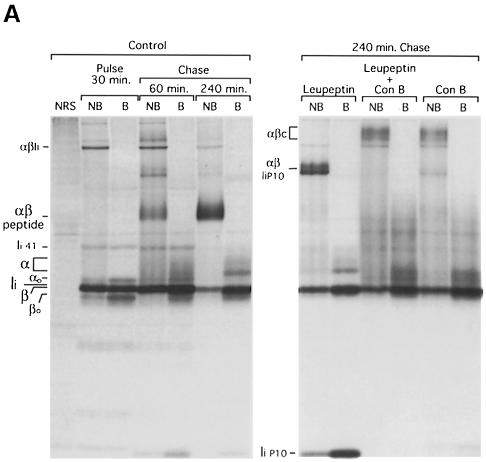
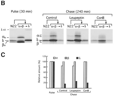
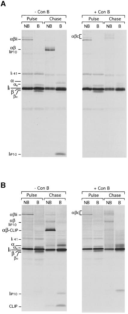
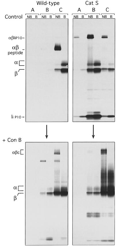
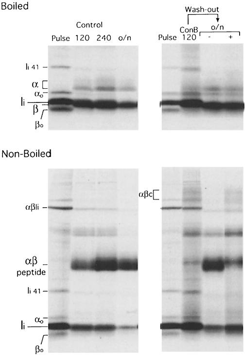
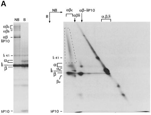
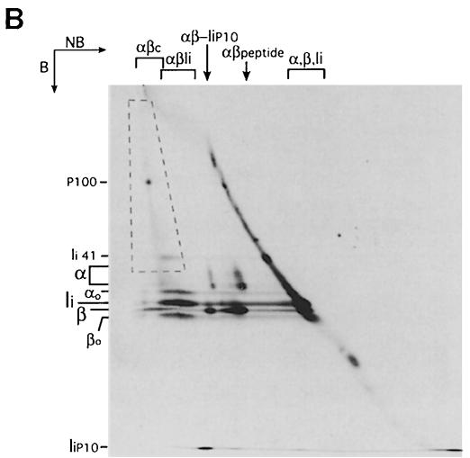
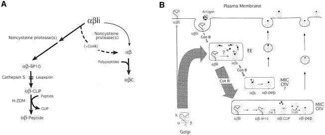
Major histocompatibility complex (MHC) class II molecules bind and present to CD4(+) T cells peptides derived from endocytosed antigens. Class II molecules associate in the endoplasmic reticulum with invariant chain (Ii), which (i) mediates the delivery of the class II-Ii complexes into the endocytic compartments where the antigenic peptides are generated; and (ii) blocks the peptide-binding site of the class II molecules until they reach their destination. Once there, Ii must be removed to allow peptide binding. The bulk of Ii-class II complexes reach late endocytic compartments where Ii is eliminated in a reaction in which the cysteine protease cathepsin S and the accessory molecule H-2DM play an essential role. Here, we here show that Ii is also eliminated in early endosomal compartments without the intervention of cysteine proteases or H-2DM. The Ii-free class II molecules generated by this alternative mechanism first bind high molecular weight polypeptides and then mature into peptide-loaded complexes.


Figures

None
Fig. 1. Ii is eliminated in ConB-treated cells without the intervention of cysteine proteases. (A) Mouse splenocytes were pulse-labeled for 30 min and chased for 60 and 240 min in the absence (control) or the presence of 1 mM leupeptin, 20 nM ConB or both drugs combined. MHC class II molecules were immunoprecipitated with mAb N22. Each immunoprecipitate was analyzed by reducing 12.5% SDS–PAGE without (NB) or after (B) boiling. The positions of immature (αo and βo) and mature (α and β) I-Ab subunits, invariant chain (Ii), the p41 form of Ii (Ii 41) (Cresswell, 1994) and the Ii degradation intermediate IiP10 are indicated. SDS-stable αβ–Ii, αβ–peptide, αβ–IiP10 and (ConB-induced) αβc complexes are also indicated. The first lane (NRS) was loaded with an immunoprecipitate obtained with normal rabbit serum plus normal mouse serum from the lysate of control cells chased for 60 min. (B) N22 immunoprecipitates obtained from pulse-labeled cells, or cells chased for 240 min without (control) or with ConB or leupeptin as in (A), were denatured by boiling in SDS. One-tenth of the sample was set apart, and the remainder used for re-immunoprecipitation with anti-I-Aα and anti-I-Aβ rabbit sera, and sequentially with a rabbit serum against the N–terminal region of Ii. The N22 immunoprecipitate and each re-immunoprecipitated sample were loaded on a reducing 12.5% SDS–polyacrylamide gel. (C) Quantitation of the re-immunoprecipitations in (B). The amount of α, β and Ii in each of the pulse, control, leupeptin and ConB sets was quantitated in a phosphorimager. To correct for differences in the total amount of sample, the values in each set were normalized relative to β. The amount of each subunit relative to the amount present in the pulse-labeled sample was calculated.
None
Fig. 2. Ii is eliminated in Cat S–/– and H-2DM–/– cells treated with ConB. Cat S–/– (A) or H-2DM–/– (B) splenocytes were pulse-labeled for 30 min and chased for 240 min in the absence or presence of 20 nM ConB, immunoprecipitated with mAb N22 and analyzed as in Figure 1. Labels are as in Figure 1. The positions of CLIP and I–Ab–CLIP that accumulate in H-2DM–/–-untreated cells are indicated (Fung-Leung et al., 1996; Martin et al., 1996; Miyazaki et al., 1996).
None
Fig. 3. ConB provokes the retention of I–Ab in early endosomes. Wild-type or Cat S–/– splenocytes were pulse-labeled for 60 min and chased for 180 min in the absence (upper panels) or presence (lower panels) of ConB. Cells were homogenized, and the post-nuclear supernatant subjected to subcellular fractionation in a two-step Percoll gradient (Castellino and Germain, 1995; Driessen et al., 1999). I–Ab was immunoprecipitated from fractions enriched in lysosomes (fraction A), late endosomes (fraction B), and early endosomes, ER, Golgi and plasma membranes (fraction C). The immunoprecipitates were analyzed by SDS–PAGE as in Figures 1 and 2.
None
Fig. 4. Reversion of ConB treatment allows formation of αβ–peptide. The panels on the left show N22 immunoprecipitates run on a 12.5% SDS–polyacrylamide gel without (lower panel) or after (upper panel) boiling, obtained from splenocytes pulse-labeled for 30 min and chased for 120 min, 240 min or overnight (o/n). In the panels on the right, the N22 immunoprecipitates were obtained from cells that had been pulse-labeled for 30 min in 20 nM ConB (pulse), then chased for 120 min in 5 nM ConB (ConB 120), and then washed and incubated overnight (o/n) in the absence (–) or presence (+) of 5 nM ConB. The high molecular weight regions of the lanes containing the boiled immunoprecipitates (upper panels) were similar to those displayed in Figures 1–3.
None
Fig. 5. The αβc complex consists of αβ dimers associated with high molecular weight polypeptides. (A) Class II molecules were immunoprecipitated from splenocytes pulsed for 30 min and chased for 240 min in the presence of ConB. A fraction of the immunoprecipitate was run on a 12.5% SDS–polyacrylamide gel without (NB) or after (B) boiling as a reference (left panel). The remainder was run first on a tubular 12.5% SDS–polyacrylamide gel without boiling. The gel was then boiled, put on top of a 10% SDS–polyacrylamide gel and run in the second dimension (right). The position of the different SDS-stable complexes and free α, β and Ii in the first dimension is indicated on top of the two-dimensional gel. The positions of immature (αo and βo) and mature (α and β) I-Ab subunits, Ii, the p41 form of Ii (Ii 41) and the Ii degradation intermediate IiP10 are indicated on the left of each gel. The dashed box encloses the area occupied by αβc-associated high molecular weight polypeptides. The percentage of polyacrylamide used in the second dimension of the two-dimensional gel (10%) is lower than in the single dimension PAGE (12.5%), causing the difference in the relative distance among polypeptides. (B) Class II molecules were immunoprecipitated from splenocytes pulse-labeled for 30 min and chased for 60 min (no drugs added). The immunoprecipitate was analyzed by two-dimensional SDS–PAGE as in (A). The position of a predominant polypeptide included in αβc is indicated (P100).
None
Fig. 6. A model for the removal of Ii from class II molecules at two different locations of the endocytic route. (A) Schematic representation of the two mechanisms of Ii elimination and the intermediates that they generate. See text for details. (B) Compartmentalization of the Cat S/H-2DM-dependent and -independent mechanisms for removal of Ii. See Dicussion for details.

Similar articles

Cited by

References

    1. Amigorena S., Webster, P., Drake, J., Newcomb, J., Cresswell, P. and Mellman, I. (1995) Invariant chain cleavage and peptide loading in major histocompatibility complex class II vesicles. J. Exp. Med., 181, 1729–1741. - PMC - PubMed
    1. Avva R.R. and Cresswell, P. (1994) In vivo and in vitro formation and dissociation of HLA–DR complexes with invariant chain-derived peptides. Immunity, 1, 763–774. - PubMed
    1. Barois N., Forquet, F. and Davoust, J. (1997) Selective modulation of the major histocompatibility complex class II antigen presentation pathway following B cell receptor ligation and protein kinase C activation. J. Biol. Chem., 272, 3641–3647. - PubMed
    1. Benaroch P., Yilla, M., Raposo, G., Ito, K., Miwa, K., Geuze, H.J. and Ploegh, H.L. (1995) How MHC class II molecules reach the endocytic pathway. EMBO J., 14, 37–49. - PMC - PubMed
    1. Bijlmakers M.J., Benaroch, P. and Ploegh, H.L. (1994) Assembly of HLA DR1 molecules translated in vitro: binding of peptide in the endoplasmic reticulum precludes association with invariant chain. EMBO J., 13, 2699–2707. - PMC - PubMed

Publication types

MeSH terms

Substances