. 2000 Mar 1;19(5):913-20.
doi: 10.1093/emboj/19.5.913.

Interaction between the tobacco mosaic virus movement protein and host cell pectin methylesterases is required for viral cell-to-cell movement

Affiliations

Interaction between the tobacco mosaic virus movement protein and host cell pectin methylesterases is required for viral cell-to-cell movement

M H Chen et al. EMBO J. .

Abstract

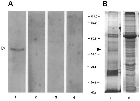
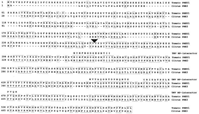
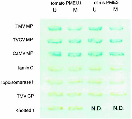
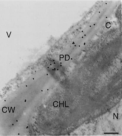
Virus-encoded movement protein (MP) mediates cell-to-cell spread of tobacco mosaic virus (TMV) through plant intercellular connections, the plasmodesmata. The molecular pathway by which TMV MP interacts with the host cell is largely unknown. To understand this process better, a cell wall-associated protein that specifically binds the viral MP was purified from tobacco leaf cell walls and identified as pectin methylesterase (PME). In addition to TMV MP, PME is recognized by MPs of turnip vein clearing virus (TVCV) and cauliflower mosaic virus (CaMV). The use of amino acid deletion mutants of TMV MP showed that its domain was necessary and sufficient for association with PME. Deletion of the PME-binding region resulted in inactivation of TMV cell-to-cell movement.


Figures

None
Fig. 1. Detection of TMV MP-interacting protein in cell wall fraction by immunorecognition of bound MP. (A) Renatured blot overlay assay. Lane 1, cell wall fraction blot incubated with MP; lane 2, cell wall fraction blot incubated with buffer alone; lane 3, empty membrane incubated with TMV MP; lane 4, soluble fraction blot incubated with TMV MP. (B) Protein content of the cell wall and soluble fractions of tobacco leaf tissue. Both lanes represent protein extract derived from 10 mg of fresh tobacco leaf tissue. Lane 1, silver-stained cell wall proteins; lane 2, Coomassie blue-stained soluble proteins. The positions of the 38 kDa TMV MP-interacting protein on the blot (open arrowhead) and on stained SDS–polyacrylamide gels (filled arrowhead) are indicated. The numbers between panels indicate molecular mass standards in kDa.
None
Fig. 2. Purification of TMV MP-interacting protein by affinity chromatography. (A) Protein fractions resolved on a 12.5% SDS–polyacrylamide gel. Protein profile was revealed by silver staining. (B) TMV MP binding on renatured protein blots. Lanes 1, total cell wall extract before loading on the column; lanes 2, flow-through fraction; lanes 3, first 0.5 ml wash fraction; lanes 4, eluted fraction. Arrowhead indicates the position of the 38 kDa TMV MP-interacting protein. The numbers on the left indicate molecular mass standards in kDa.
None
Fig. 3. Amino acid sequence of three peptides derived from the purified TMV MP-interacting protein and its alignment with full-length PME sequences from tomato (DDBJ/EMBL/GenBank accession No. U49330) and citrus (Valencia orange, DDBJ/EMBL/GenBank accession No. U82977). Alignment was performed by the clustal algorithm. Regions of identity between tomato and citrus PMEs are indicated by boxes, gaps introduced for alignment are indicated by dashes. The arrowhead indicates the N–terminus of the mature form of PME (according to Gaffe et al., 1997; Nairn et al., 1998).
None
Fig. 4. Interaction of PME with plant viral MPs in the yeast two-hybrid system. Yeast cells expressing the combination of interacting proteins indicated were grown on tryptophan–leucine double dropout medium and analyzed for β–galactosidase activity. N.D., not done. U, unprocessed form of PME; M, mature form of PME.
None
Fig. 5. Immunohistochemical detection of PME in cell walls of tobacco leaf mesophyll cells. CW, cell wall; PD, plasmodesma; N, nucleus, CHL, chloroplast; V, vacuole; C, cytoplasm. Bar, 0.5 μm.
None
Fig. 6. Analysis of MP deletion mutants for their ability to bind tobacco PME on renatured blot overlays. (A) Summary of the deletion mutants tested. Ovals indicate retained amino acids; lines indicate deleted amino acids. The numbers in the scale refer to amino acids of the wild-type (wt) TMV MP (Goelet et al., 1982). (B) Renatured blot overlay binding assay using tobacco cell wall fractions. Each lane is designated with the TMV MP derivative whose binding was tested.
None
Fig. 7. Analysis of MP deletion mutants for their ability to bind tomato PME in a yeast two-hybrid system. For description of TMV MP deletion mutants see Figure 6. TMV MP wt, full-length MP; TMV MP130–185, MP fragment composed of amino acid residues 130–185. U, unprocessed form of PME; M, mature form of PME.

Similar articles

Cited by

References

    1. Atkins D., Hull, R., Wells, B., Roberts, K., Moore, P. and Beachy, R.N. (1991) The tobacco mosaic virus 30 K movement protein in transgenic tobacco plants is localized to plasmodesmata. J. Gen. Virol., 72, 209–211. - PubMed
    1. Bartel P., Chien, C., Sternglanz, R. and Fields, S. (1993) Elimination of false positives that arise in using the two-hybrid system. BioTechniques, 14, 920–924. - PubMed
    1. Carrington J.C., Kassachau, K.D., Mahajan, S.K. and Schaad, M.C. (1996) Cell-to-cell and long-distance transport of viruses in plants. Plant Cell, 8, 1669–1681. - PMC - PubMed
    1. Citovsky V. and Zambryski, P. (1993) Transport of nucleic acids through membrane channels: Snaking through small holes. Annu. Rev. Microbiol., 47, 167–197. - PubMed
    1. Citovsky V. and Zambryski, P. (1995) Transport of protein–nucleic acid complexes within and between plant cells. Membr. Protein Transp., 1, 39–57.

Publication types