. 2000 Mar 1;19(5):979-88.
doi: 10.1093/emboj/19.5.979.

Identification of a pocket in the PDK1 kinase domain that interacts with PIF and the C-terminal residues of PKA

Affiliations

Identification of a pocket in the PDK1 kinase domain that interacts with PIF and the C-terminal residues of PKA

R M Biondi et al. EMBO J. .

Abstract

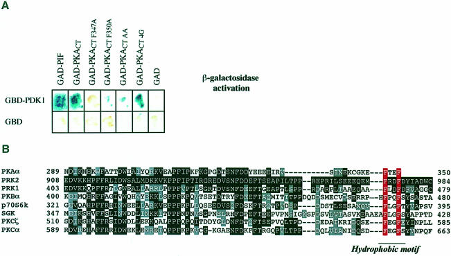
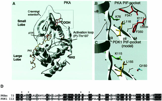
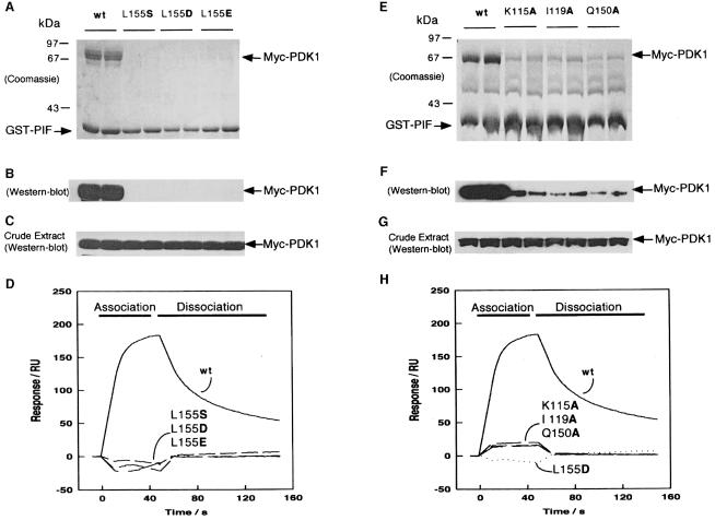
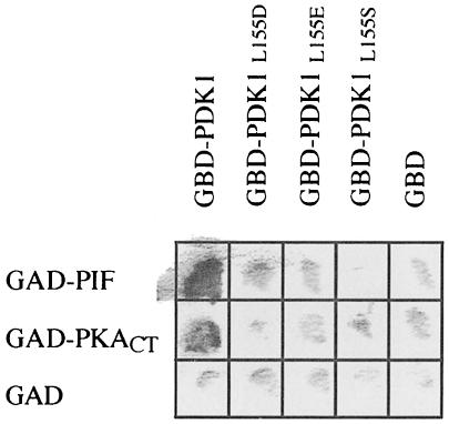
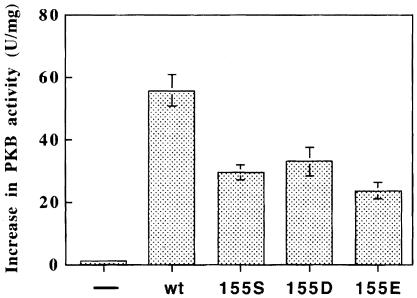
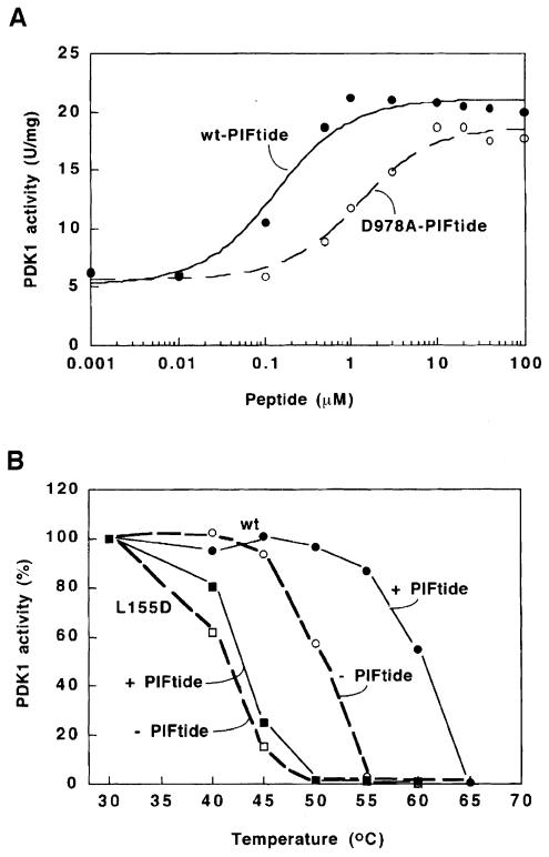
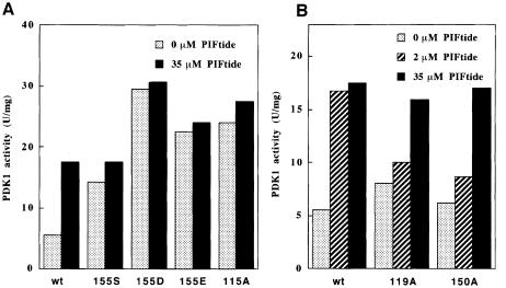
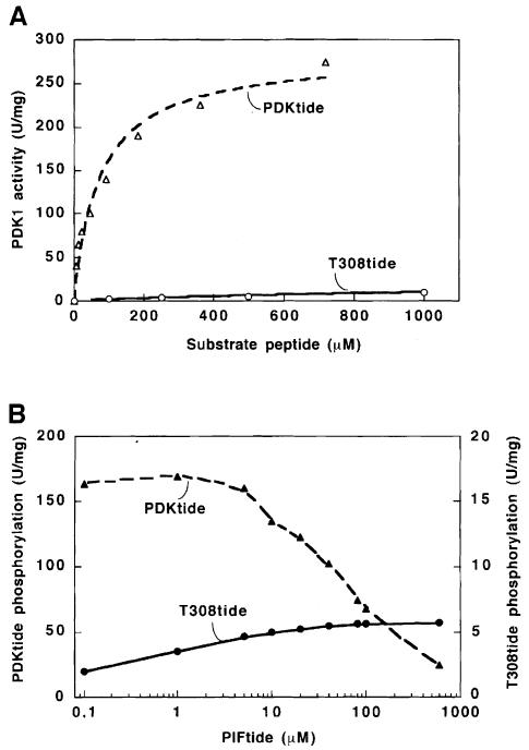
The 3-phosphoinositide-dependent protein kinase-1 (PDK1) phosphorylates and activates a number of protein kinases of the AGC subfamily. The kinase domain of PDK1 interacts with a region of protein kinase C-related kinase-2 (PRK2), termed the PDK1-interacting fragment (PIF), through a hydrophobic motif. Here we identify a hydrophobic pocket in the small lobe of the PDK1 kinase domain, separate from the ATP- and substrate-binding sites, that interacts with PIF. Mutation of residues predicted to form part of this hydrophobic pocket either abolished or significantly diminished the affinity of PDK1 for PIF. PIF increased the rate at which PDK1 phosphorylated a synthetic dodecapeptide (T308tide), corresponding to the sequences surrounding the PDK1 phosphorylation site of PKB. This peptide is a poor substrate for PDK1, but a peptide comprising T308tide fused to the PDK1-binding motif of PIF was a vastly superior substrate for PDK1. Our results suggest that the PIF-binding pocket on the kinase domain of PDK1 acts as a 'docking site', enabling it to interact with and enhance the phosphorylation of its substrates.


Figures

None
Fig. 1. Two-hybrid interaction of PDK1 and wild-type and mutant C–terminal fragment of PKA. (A) The Y190 yeast strain was transformed with vectors expressing PDK1 fused to the Gal4 DNA-binding domain (GBD), together with vectors encoding either the C–terminal 26 residues of PIF or the wild-type, or the indicated mutants of a C–terminal fragment of PKA (PKACT residues 129–350) fused to a Gal4 activation domain (GAD). As a control, yeast were also co-transformed with the GBD domain alone and the GAD domain alone. The yeast were grown overnight at 30°C and β–galactosidase filter lift assays performed at 30°C for 4 h. An interaction between GBD–PDK1 and GAD–PKACT induces the expression of β–galactosidase, which is detected as a blue colour in the filter lift assay. (B) Alignment of the amino acid sequence of the C–terminal 77 amino acids of PKA with the equivalent region of the AGC subfamily kinases indicated. Identical residues are denoted by white letters on a black background, and similar residues by grey boxes. The aromatic residues in the hydrophobic motif are indicated in red.
None
Fig. 2. C–terminal Phe-Xaa-Xaa-Phe residues of PKA interact with a hydrophobic pocket on the PKA kinase domain, predicted to be conserved in PDK1. (A) Ribbon structure of the PKA–PKI–ATP ternary complex (Zheng et al., 1993); PKI is shown in yellow, and the ATP molecule is highlighted. The C–terminal Phe347 and Phe350 are shown in red. The position of phospho-Thr197 (the PDK1 phosphorylation site) in the T-loop is indicated. (B) Detailed structure of the hydrophobic pocket on the kinase domain of PKA that interacts with the C–terminal Phe-Xaa-Xaa-Phe residues of PKA. Lys76 (equivalent of Lys115 in PDK1) is shown in green, Leu116 (equivalent of Leu155 in PDK1) is shown in yellow, Phe347 and Phe350 in red, and certain amine residues are in blue. (C) The structure of the PDK1 kinase domain was modelled as described in Materials and methods. The region of PDK1 equivalent to the hydrophobic pocket of PKA termed the PIF-binding pocket is shown. The residues predicted to be involved in binding to PIF are highlighted, including the amide residue of Q150. (D) Alignment of the amino acid residues of PDK1 around the PIF-binding pocket and the equivalent region of PKA. Identical residues are denoted by white letters on a black background, and similar residues by grey boxes. Residues on PKA that interact with the C–terminal Phe-Xaa-Xaa-Phe motif are marked with an asterisk.
None
Fig. 3. Effect of mutation of conserved residues in the PIF-binding pocket of PDK1 on the ability of PDK1 to interact with PIF. 293 cells were transiently transfected with DNA constructs expressing GST–PIF and either wild-type Myc-PDK1 or the indicated mutants of PDK1. At 36 h post-transfection, the cells were lysed and GST–PIF purified by affinity chromatography on glutathione–Sepharose beads. A 2 μg aliquot of each protein was electrophoresed on a 10% SDS–polyacrylamide gel and either stained with Coomassie Blue (A and E) or immunoblotted using an anti-Myc antibody to detect Myc-PDK1 (B and F). To establish that the wild-type PDK1 and mutant proteins were expressed at a similar level, 10 μg of total cell lysate was electrophoresed on a 10% SDS–polyacrylamide gel and immunoblotted using anti-Myc antibodies (C and G). Duplicates of each condition are shown. Similar results were obtained in three to five separate experiments. (D and H) Surface plasmon resonance measurements were carried out on a BiaCore instrument as described in Materials and methods to measure the interaction of wild-type and mutant GST–PDK1 preparations with the 24 residue synthetic peptide whose sequence encompasses the PDK1-binding site on PIF termed PIFtide (Balendran et al., 1999a). PIFtide was immobilized on an SA SensorChip, and wild-type (wt) or the indicated mutants of PDK1 were injected at a concentration of 40 nM. All data are single determinations from a representative experiment that was repeated at least three times with similar results. For clarity, the bulk refractive index changes associated with the first and last 10 s of the injection have been removed.
None
Fig. 4. Leu155 mutants of PDK1 do not interact with either PIF or the C–terminal fragment of PKA in the two-hybrid system. The Y190 yeast strain was transformed with vectors expressing the wild-type PDK1 or the indicated mutants of PDK1 fused to the Gal4 DNA-binding domain (GBD) together with vectors encoding the expression of either the 26 C–terminal residues of PRK2 (PIF) or the C–terminal fragment of PKA (PKACT residues 129–350) fused to a Gal4 activation domain (GAD). As a control, yeast were also co-transformed with vectors expressing the GAD and GBD domains only. The yeast were grown overnight at 30°C and β–galactosidase filter lift assays performed at 30°C for 4 h. An interaction between GBD–PDK1 and either GAD–PIF or GAD–PKACT induces the expression of β–galactosidase, which is detected as a blue colour (shown as black in the figure) in the filter lift assay.
None
Fig. 5. Phosphorylation of Thr308 of PKB by wild-type and PIF-binding pocket mutants of PDK1. Wild-type or mutant forms of GST–PDK1 were expressed in 293 cells and purified by affinity chromatography on glutathione–Sepharose beads. Each GST fusion protein (0.2 ng) was incubated for 30 min at 30°C with GST–S473D-PKBα and MgATP in the presence or absence of phospholipid vesicles containing 100 μM phosphatidycholine, 100 μM phosphatidylserine and 10 μM sn-1-stearoyl-2-arachidonoyl-d-PtdIns(3,4,5)P3, and the increase in specific activity of GST–S473D-PKBα was determined relative to a control incubation in which the PDK1 was omitted (average of six determinations, three independent experiments). The basal activity of GST–S473D-PKBα was 1.5 U/mg. Under the conditions used, it was verified that the activation of GST–473D-PKBα was proportional to the amount of PDK1 added to the assay (data not shown). ‘–’ indicates that PDK1 was omitted.
None
Fig. 6. PDK1 is activated and stabilized through its interaction with PIFtide. (A) GST–PDK1 activity was measured in the presence of increasing concentrations of wild-type (wt) PIFtide (•) or a mutant D978A-PIFtide (○), using the synthetic peptide substrate termed T308tide, as described in Materials and methods. The data were fitted to a hyperbola using the Kaleidagraph software. The concentration needed to obtain 50% activation of PDK1 was 0.14 μM for wt-PIFtide and 1.1 μM for D978A-PIFtide. The assay shown was performed in triplicate and there was <5% difference between each assay. Similar results were obtained in two further experiments. (B) The wild-type GST–PDK1 (circles) or the L155D mutant of GST–PDK1 (squares) was incubated in the presence (closed symbols) or absence (open symbols) of 100 μM PIFtide and then heated for 2 min at the indicated temperatures, rapidly brought to 0°C (see Materials and methods), and 2 min later assayed at 30°C for 10 min using T308tide as substrate. The activity of PDK1 obtained by incubation at 30°C was taken as 100%. The assay shown was performed in duplicate with similar results obtained in two separate experiments.
None
Fig. 7. Effect of PIFtide on PDK1 PIF pocket mutants. Wild-type and the indicated mutants of GST–PDK1 were assayed with T308tide either in the absence (dotted bars) or the presence of 2 mM PIFtide (dashed bars) or 35 μM PIFtide (filled bars). Under the conditions used, the phosphorylation of T308tide by PDK1was linear with time (data not shown). (A) The PDK1 mutants that are activated in the absence of PIFtide and (B) those mutants that are activated by high concentrations of PIFtide. The assay was performed in triplicate with <10% difference between triplicate samples. Similar results were obtained in three separate experiments.
None
Fig. 8. PDKtide is vastly superior to T308tide as a substrate for PDK1 because it interacts with the PIF-binding pocket of PDK1. (A) His-PDK1 was assayed for activity using as substrate the indicated concentration of either PDKtide (▵) or T308tide (○). (B) His-PDK1 was assayed for activity in the presence of PDKtide (25 μM, ▴) or T308tide (100 μM, •) in the presence of the indicated concentrations of PIFtide. The assay was performed in triplicate with <5% difference between the triplicate samples. Similar results were obtained in three separate experiments

Similar articles

Cited by

References

    1. Alessi D.R. and Downes, C.P. (1998) The role of PI 3-kinase in insulin action. Biochim. Biophys. Acta, 1436, 151–164. - PubMed
    1. Alessi D.R., Cohen, P., Ashworth, A., Cowley, S., Leevers, S.L. and Marshall, C.J. (1994) Assay and expression of mitogen activated protein kinase, MAP kinase and Raf. Methods Enzymol., 255, 279–290. - PubMed
    1. Alessi D.R., Andjelkovic, M., Caudwell, B., Cron, P., Morrice, N., Cohen, P. and Hemmings, B.A. (1996) Mechanism of activation of protein kinase B by insulin and IGF-1. EMBO J., 15, 6541–6551. - PMC - PubMed
    1. Alessi D.R., et al. (1997a) 3-Phosphoinositide-dependent protein kinase-1 (PDK1): structural and functional homology with the Drosophila DSTPK61 kinase. Curr. Biol., 7, 776–789. - PubMed
    1. Alessi D.R., James, S.R., Downes, C.P., Holmes, A.B., Gaffney, P.R., Reese, C.B. and Cohen, P. (1997b) Characterization of a 3-phospho– inositide-dependent protein kinase which phosphorylates and activates protein kinase Bα. Curr. Biol., 7, 261–269. - PubMed

Publication types

MeSH terms

Substances