. 2000 Mar 1;19(5):1087-97.
doi: 10.1093/emboj/19.5.1087.

Functional interaction between RAFT1/FRAP/mTOR and protein kinase cdelta in the regulation of cap-dependent initiation of translation

Affiliations

Functional interaction between RAFT1/FRAP/mTOR and protein kinase cdelta in the regulation of cap-dependent initiation of translation

V Kumar et al. EMBO J. .

Abstract

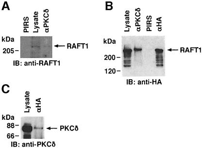
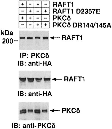
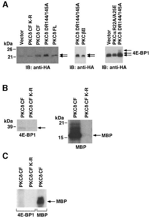
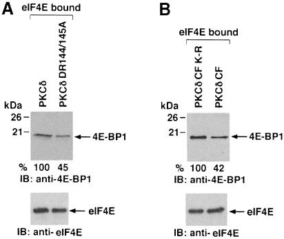
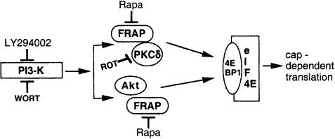
Hormones and growth factors induce protein translation in part by phosphorylation of the eukaryotic initiation factor 4E (eIF4E) binding protein 1 (4E-BP1). The rapamycin and FK506-binding protein (FKBP)-target 1 (RAFT1, also known as FRAP) is a mammalian homolog of the Saccharomyces cerevisiae target of rapamycin proteins (mTOR) that regulates 4E-BP1. However, the molecular mechanisms involved in growth factor-initiated phosphorylation of 4E-BP1 are not well understood. Here we demonstrate that protein kinase Cdelta (PKCdelta) associates with RAFT1 and that PKCdelta is required for the phosphorylation and inactivation of 4E-BP1. PKCdelta-mediated phosphorylation of 4E-BP1 is wortmannin resistant but rapamycin sensitive. As shown for serum, phosphorylation of 4E-BP1 by PKCdelta inhibits the interaction between 4E-BP1 and eIF4E and stimulates cap-dependent translation. Moreover, a dominant-negative mutant of PKCdelta inhibits serum-induced phosphorylation of 4E-BP1. These findings demonstrate that PKCdelta associates with RAFT1 and thereby regulates phosphorylation of 4E-BP1 and cap-dependent initiation of protein translation.


Figures

None
Fig. 1. Association of PKCδ with RAFT1. (A) Total lysates from 293T cells were subjected to immunoprecipitation with anti-PKCδ or pre-immune rabbit serum (PIRS). The precipitates and total lysate were separated by SDS–PAGE and analyzed by immunoblotting with anti-RAFT1. (B) 293T cells were transiently transfected with HA-RAFT1. Total lysates were subjected to immunoprecipitation with anti-PKCδ, anti-HA or PIRS. The precipitates and the lysates were analyzed by immunoblotting with anti-HA. (C) 293T cells were transiently transfected with HA-RAFT1. Lysates were subjected to immunoprecipitation with anti-HA. The precipitates and lysates were analyzed by immunoblotting with anti-PKCδ.
None
Fig. 2. Kinase activities of PKCδ and RAFT1 are not required for their association. 293T cells were transiently transfected with PKCδ FL or PKCδ DR144/145A with HA-RAFT1 or HA-RAFT1 D2357E. Lysates were subjected to immunoprecipitation with anti-PKCδ and the precipitates were analyzed by immunoblotting with anti-HA (top panel). Anti-HA immunoprecipitates were analyzed by immunoblotting with anti-HA (middle panel). Lysates were also analyzed by immunoblotting with anti-PKCδ (bottom panel).
None
Fig. 3. PKCδ induces phosphorylation of 4E-BP1. (A) 293T cells were transiently transfected with HA-4E-BP1 and vector, PKCδ FL or PKCδ DR144/145A. Cells were serum starved for 48 h and lysates were resolved by SDS–PAGE and analyzed by immunoblotting with anti-HA. 293T cells were transiently cotransfected with HA-4E-BP1 and vector, PKCδ CF or PKCδ CF(K–R). Cells were serum starved for 48 h. Lysates were resolved by SDS–PAGE and analyzed by immunoblotting with anti-HA (left panel). 293T cells were transiently transfected with HA-4E-BP1 and PKCβII or PKCδ DR144/145A (middle panel). 293T cells were transfected with HA-4E-BP1 and vector, PKCα R22A/A25E or PKCδ DR144/145A (right panel). Cells were serum starved for 48 h. Lysates were resolved by SDS–PAGE and analyzed by immunoblotting with anti-HA. (B) GST–PKCδ CF or GST–PKCδ CF(K–R) fusion proteins were incubated with purified GST–4E-BP1 in the presence of [γ-32P]ATP for 15 min at 30°C (left panel). GST–PKCδ CF or GST–PKCδ CF(K–R) were also separately incubated with MBP (right panel). The reaction products were resolved by SDS–PAGE and analyzed by autoradiography. (C) 293 cells were serum starved for 36 h. Lysates were incubated with m7GTP resin and the precipitates were incubated with PKCδ CF in the presence of [γ-32P]ATP and MBP and analyzed by SDS–PAGE and autoradiography.
None
Fig. 4. PKCδ inhibits the interaction of eIF4E with 4E-BP1. (A) 293T cells were transiently cotransfected with PKCδ FL or PKCδ DR144/145A and serum starved for 48 h. Lysates were incubated with m7GTP–agarose beads for 45 min at 4°C. After incubation, the proteins were resolved by 15% SDS–PAGE and analyzed by immunoblotting with anti-4E-BP1 (upper panel) or anti-eIF4E (bottom panel). (B) 293T cells were transiently transfected with PKCδ CF or PKCδ CF(K–R) and serum starved for 48 h. Lysates were incubated with m7GTP–agarose beads for 45 min at 4°C. After incubation, the proteins were resolved by 15% SDS–PAGE and analyzed by immuno- blotting with anti-4E-BP1 (upper panel) or anti-eIF4E (bottom panel).
None
Fig. 5. Effect of PKCδ on cap-dependent protein translation. (A) 293T cells were transiently cotransfected with pcDNA3-LUC-pol-CAT (2 μg) and empty vector, PKCβII or PKCδ DR144/145A. Cells were serum starved for 48 h and lysates were analyzed for luciferase activity. Activity is expressed as percentage control (mean ± SD of three independent transfections). 293T cells were cotransfected with pcDNA3-LUC-pol-CAT (2 μg) and vector or PKCδ CF. Following serum starvation for 48 h, total cell lysates were analyzed for luciferase activity. (B) 293T cells were transiently cotransfected with pcDNA3-LUC-pol-CAT (2 μg) and vector (10 μg), PKCα R22A/A25E (10 μg) or PKCδ DR144/145A (10 μg). Following serum starvation for 48 h, lysates were assayed for luciferase activity. (C) 293T cells were transiently transfected with vector, pcDNA3-LUC-pol-CAT and the amounts of PKCδ DR144/145A indicated. Lysates were assayed for luciferase activity. Luciferase activity is expressed as percentage control (mean ± SD of four independent transfections). (D) Antisense GAPDH and luciferase probes (lanes 1 and 2) were tested against RNA from PKCδ CF (lane 3), PKCδ CF(K–R) (lane 4), PKCδ FL (lane 5), PKCδ DR144/145A (lane 6) or mock (lane 7) transfected cells. Positions of FL probes and protected fragments are indicated on the right.
None
Fig. 6. PKCδ-mediated increase in cap-dependent protein translation requires activity of RAFT1 and is resistant to wortmannin and sensitive to rapamycin. (A) 293 cells were cotransfected with pcDNA3-LUC-pol-CAT (2 μg) and vector (10 μg) (bar 1), RAFT1 (10 μg) (bar 2), RAFT1 D2357E (10 μg) (bar 3), PKCδ DR144/145A (10 μg) (bar 4), PKCδ DR144/145A + RAFT1 (bar 5) or PKCδ DR144/145A + RAFT1 D2357E (bar 6). Cells were serum starved for 48 h and lysates were assayed for luciferase activity. Luciferase activity is expressed as percentage control (mean ± SD of three independent transfections). 293 cells were cotransfected with pcDNA3-LUC-pol-CAT (2 μg) and vector or PKCδ CF in the presence or absence of HA-RAFT1 D2357E. Cells were serum starved for 48 h and lysates were assayed for luciferase activity. (B) Left panel: 293 cells were transfected with HA-4E-BP1 with vector (lane 1); PKCδ DR144/145A + RAFT1 wild type (lane 2); PKCδ DR144/145A (lane 3) and PKCδ DR144/145A + RAFT1 D2357E (lane 4). Lysates were incubated with m7GTP resin and the precipitates were analyzed by immunoblotting with anti-HA (left panel). Right panel: 293 cells were also separately cotransfected with HA-4E-BP1 and PKCδ CF + RAFT1 D–E (lane 1) or PKCδ CF + RAFT1 wild type (lane 2). Lysates were incubated with m7GTP resin and the precipitates were analyzed by immunoblotting with anti-HA. (C) 293 cells were cotransfected with pcDNA3-LUC-pol-CAT (2 μg) and PKCδ DR144/145A mutant or PKCδ CF. After transfection, cells were serum starved for 48 h and then treated with 20 ng/ml rapamycin. Lysates were assayed for luciferase activity. (D) 293 cells were cotransfected with HA-4E-BP1 and PKCδ DR144/145A. Cells were also separately transfected with vector and HA-4E-BP1. Cells were serum starved for 48 h after transfection and then treated with buffer (lane 1), 20 ng/ml rapamycin (lane 2) or 200 nM wortmannin (lane 3). Lysates were analyzed by immunoblotting with anti-HA (top panel). The results are expressed as percentage inhibition (mean ± SD) of three independent experiments (bottom panel).
None
Fig. 7. Role of PKCδ in cap-dependent protein translation in vivo. (A) MCF-7/GFP and MCF-7/GFP–PKCδ-RD cells were transfected with pcDNA3-LUC-pol-CAT. Cells were serum starved for 36 h and stimulated with serum in the presence or absence of 50 ng/ml rapamycin for an additional 12 h. Lysates were assayed for luciferase activity. Luciferase activity is expressed as percentage control (mean ± SD of three independent experiments). Total cell lysates from MCF-7/GFP and MCF-7/GFP–PKCδ-RD cells were also analyzed by immunoblotting with anti-GFP (inset). (B) MCF-7/GFP and MCF-7/GFP–PKCδ-RD cells were transfected with HA-4E-BP1. Cells were serum starved for 36 h and stimulated with serum for 45 and 90 min. Lysates were analyzed by immunoblotting with anti-HA (upper panel). The results are expressed as percentage 4E-BP1 phosphorylation (mean ± SD) of two independent experiments (lower panel). (C) 293 cells were transfected with vector or GFP–PKCδ-RD. Total cell lysates were subjected to immunoprecipitation with anti-PKCδ and the precipitates were incubated with MBP in the presence of [γ-32P]ATP. The reaction products were analyzed by SDS–PAGE and auto- radiography (top panel). Anti-PKCδ immunoprecipitates were analyzed by immunoblotting with anti-PKCδ (middle panel). Lysates were also analyzed by immunoblotting with anti-GFP (bottom panel).
None
Fig. 8. PKCδ inhibitor, rottlerin, inhibits serum-induced phosphoryl- ation of 4E-BP1. (A) 293 cells were serum starved for 36 h and stimulated with serum in the presence or absence of PKCδ inhibitor, rottlerin (Rot). Lysates were analyzed by immunoblotting with anti-4E-BP1. (B) 293 cells were serum starved for 36 h and then stimulated with serum in the presence or absence of rottlerin. Lysates were incubated with m7GTP–agarose beads for 45 min at 4°C. After incubation, the bound proteins were resolved by 15% SDS–PAGE and analyzed by immunoblotting with anti-4E-BP1. (C) 293 cells were serum starved for 36 h and stimulated with serum in the presence or absence of rottlerin (Rot). Total cell lysates were subjected to incubation with anti-PKCδ (filled bars) or anti-Akt (hatched bars). Precipitates were incubated in kinase buffer containing [γ-32P]ATP and MBP. The reaction products were analyzed by SDS–PAGE and autoradiography. The results are expressed as PKCδ activity (mean ± SD) of two independent experiments.
None
Fig. 9. Schematic model depicting the role of the mTOR/FRAP/RAFT1–PKCδ complex in 4E-BP1-mediated, cap-dependent translation. PI3-K acts upstream to the mTOR/FRAP–Akt pathway (Gingras et al., 1998). The present results demonstrate that overexpression of active PKCδ DR144/145A is associated with induction in phosphorylation of 4E-BP1 and that this effect is insensitive to wortmannin. These findings indicate that the mTOR/FRAP–PKCδ complex lies downstream to PI3-K. Moreover, there is no detectable interaction between Akt and PKCδ (data not shown). Our results and those from others (Gingras et al., 1998) indicate that FRAP/mTOR functions as a scaffolding protein through which multiple upstream effectors converge and thereby initiate cap-dependent translation. [PKCδ–mTOR/FRAP; Akt–mTOR/FRAP]→→4E-BP1.

Similar articles

Cited by

References

    1. Abraham R.T. (1996) Phosphoinositol 3-kinase related kinases. Curr. Opin. Immunol., 8, 412–418. - PubMed
    1. Alessi D.R., et al. (1997) Phosphoinositide-dependent protein kinase-1 (PDK1): structural and functional homology with the Drosophila DSTPK61 kinase. Curr. Biol., 7, 776–789. - PubMed
    1. Barbet N., Schneider, U., Helliwell, S.B., Stansfield, I., Tuite, M.F. and Hall, M.N. (1996) TOR controls translation initiation and early G1 progression in yeast. Mol. Cell. Biol., 7, 25–42. - PMC - PubMed
    1. Belham C., Wu, S. and Avruch, J. (1999) Intracellular signaling: PDK1–a kinase at the hub of things. Curr. Biol., 9, R93–R96. - PubMed
    1. Beretta L., Gingras, A.C., Svitkin, Y.V., Hall, A.N. and Sonenberg, N. (1996) Rapamycin blocks the phosphorylation of 4E-BP1 and inhibits cap-dependent initiation of translation. EMBO J., 15, 658–664. - PMC - PubMed

Publication types

MeSH terms