. 2000 Mar 1;19(5):1157-66.
doi: 10.1093/emboj/19.5.1157.

UV damage causes uncontrolled DNA breakage in cells from patients with combined features of XP-D and Cockayne syndrome

Affiliations

UV damage causes uncontrolled DNA breakage in cells from patients with combined features of XP-D and Cockayne syndrome

M Berneburg et al. EMBO J. .

Abstract

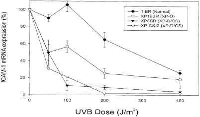
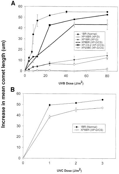
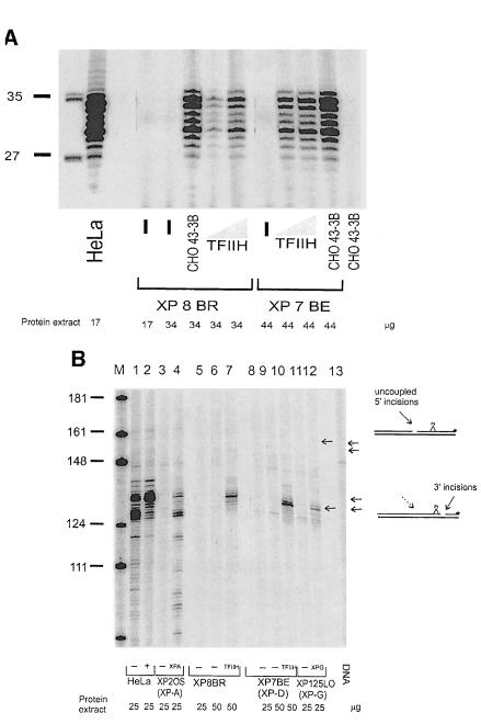
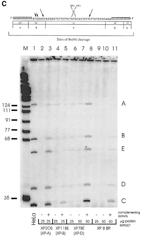
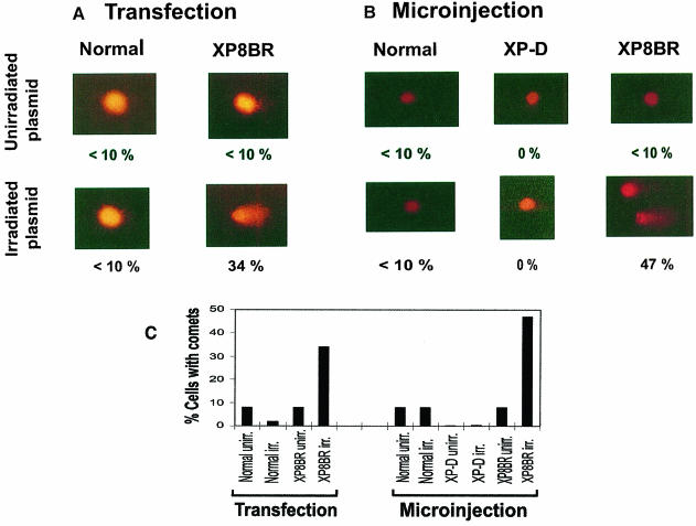
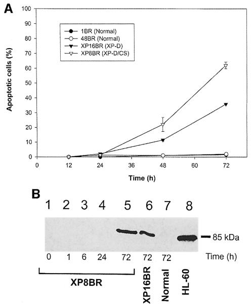
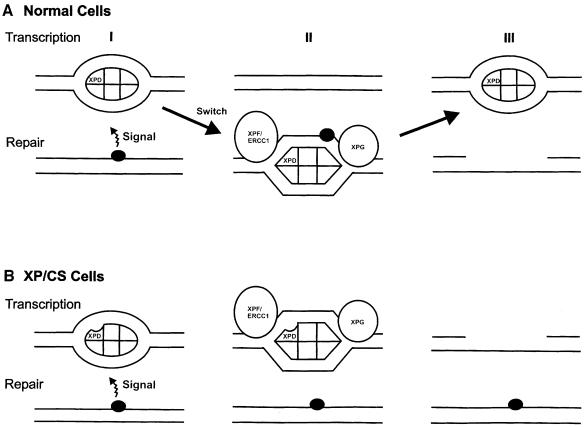
Nucleotide excision repair (NER) removes damage from DNA in a tightly regulated multiprotein process. Defects in NER result in three different human disorders, xeroderma pigmentosum (XP), trichothiodystrophy (TTD) and Cockayne syndrome (CS). Two cases with the combined features of XP and CS have been assigned to the XP-D complementation group. Despite their extreme UV sensitivity, these cells appeared to incise their DNA as efficiently as normal cells in response to UV damage. These incisions were, however, uncoupled from the rest of the repair process. Using cell-free extracts, we were unable to detect any incision activity in the neighbourhood of the damage. When irradiated plasmids were introduced into unirradiated XP-D/CS cells, the ectopically introduced damage triggered the induction of breaks in the undamaged genomic DNA. XP-D/CS cells thus have a unique response to sensing UV damage, which results in the introduction of breaks into the DNA at sites distant from the damage. We propose that it is these spurious breaks that are responsible for the extreme UV sensitivity of these cells.


Figures

None
Fig. 1. Inhibition of ICAM–1 expression by UVB. Normal, XP–D (XP16BR) or XP–D/CS fibroblasts were exposed to different doses of UVB irradiation and then treated with interferon–γ. Four hours later the levels of ICAM–1 mRNA were measured by semi-quantitative RT–PCR. Error bars represent the SEMs of three experiments.
None
Fig. 2. Accumulation of incised intermediates after UV irradiation of normal and XP–D/CS cells. Fibroblasts were irradiated with different doses of UVB (A) or UVC (B) and incubated for 1 h in the presence of HU and ara–C. Incised intermediates were measured using the comet assay. The average increase in length of the comet tails is plotted as a function of UV dose.
None
Fig. 3. Accumulation of incised intermediates in the absence of repair synthesis inhibitors after UV irradiation. Non-proliferating cells were (A) irradiated with different doses of UVB and incubated for 1 h, or (B) irradiated with 30 J/m2 UVB and incubated for different times prior to analysis of the DNA using the comet assay.
None
Fig. 4. Absence of NER in cell-free extracts of XP–D/CS cells. (A) Cell-free extracts of HeLa, XP7BE or XP8BR cells were incubated with plasmid DNA containing a single cisplatin lesion. Excised fragments were directly end-labelled with [32P]dCTP and separated on polyacrylamide gels. Different amounts of cell extract protein were used as indicated, and in some samples they were supplemented either with TFIIH or with 30 μg of protein extract from the NER-deficient Chinese hamster ovary cell line 43-3B. (B) To analyse incision activity of cell extracts, the amounts of extract protein indicated were incubated with substrate and a DNA fragment containing the lesion was isolated by restriction digestion and 3′-end-labelled. M, size marker. (C) Repair synthesis. Substrate was incubated with extracts and triphosphates including radioactive dCTP to label repair patches. The DNA was then digested with BstNI and the products separated on polyacrylamide gels.
None
Fig. 5. Introduction of irradiated plasmids causes breakage of genomic DNA in XP–D/CS cells. (A) Transfection. SV40-transformed 1BR.neo or XP8BR.neo cells were cotransfected with unirradiated or UV-irradiated pcDNA3.1 and pHook–1. Twenty-four hours later, the cultures were harvested and cells expressing the pHook–1 plasmid were separated with hapten-linked magnetic beads, embedded in agarose and the genomic DNA analysed using the comet assay. Typical cells are shown after electrophoresis in the comet assay, together with the percentage of cells with comet tails >20 μm. (B) Microinjection. Normal, XP–D (XP3NE) or XP8BR cells in a marked area on a dish were microinjected with UV-irradiated pcDNA3.1 and incubated for 6 h, before harvesting and analysis using the comet assay. Details as in (A). (C) The data are summarized as a percentage of cells with comet tails >20 μm under different conditions.
None
Fig. 6. Apoptosis in normal and XP8BR cells after UVB irradiation. Cells were irradiated with 100 J/m2 UVB and at various times after irradiation apoptosis was measured either using the Apoptag kit (A) or by the cleavage of PARP (B). In the latter experiments, sonicated cell lysates were electrophoresed in SDS–PAGE gels and the gels analysed by Western blotting using an antibody specific for the 85 kDa cleavage product of PARP.
None
Fig. 7. Model for the effects of the defect in XP–D/CS cells. (A) In normal cells (I), transcription initiation utilizes TFIIH in its transcription mode (depicted as an ellipse). A signal from a DNA damaged site recruits TFIIH and converts it into repair mode (depicted as a hexagon) (II) and NER takes place. After incision, TFIIH reverts to its transcriptional configuration (III). (B) In XP–D/CS cells, the signal from the damaged site converts TFIIH into its repair conformation (II), but is unable to recruit it to the site of the damage. Nicks are therefore generated at sites of transcription initiation, instead of at repair sites (III).

References

    1. Aboussekhra A., et al. (1995) Mammalian DNA nucleotide excision repair reconstituted with purified components. Cell, 80, 859–868. - PubMed
    1. Ahrens C., et al. (1997) Photocarcinogenesis correlates with immunosuppression in DNA-repair-defective individuals. Proc. Natl Acad. Sci. USA, 94, 6837–6841. - PMC - PubMed
    1. Berneburg M., Clingen, P.H., Harcourt, S.A., Lowe, J.E., Taylor, E.M., Green, M.H.L., Krutmann, J., Arlett, C.F. and Lehmann, A.R. (2000) The cancer-free phenotype in trichothiodystrophy is unrelated to its repair defect. Cancer Res., 60, 431–438. - PubMed
    1. Botta E., Nardo, T., Broughton, B.C., Marinoni, S., Lehmann, A.R. and Stefanini, M. (1998) Analysis of mutations in the XPD gene in Italian patients with trichothiodystrophy: site of mutation correlates with repair deficiency but gene dosage appears to determine clinical severity. Am. J. Hum. Genet., 63, 190–196. - PMC - PubMed
    1. Broughton B.C., et al. (1995) Molecular and cellular analysis of the DNA repair defect in a patient in xeroderma pigmentosum complementation group D with the clinical features of xeroderma pigmentosum and Cockayne syndrome. Am. J. Hum. Genet., 56, 167–174. - PMC - PubMed

Publication types