. 2000 Mar;38(3):965-70.
doi: 10.1128/JCM.38.3.965-970.2000.

Genotyping of Mycoplasma pneumoniae clinical isolates reveals eight P1 subtypes within two genomic groups

Affiliations

Genotyping of Mycoplasma pneumoniae clinical isolates reveals eight P1 subtypes within two genomic groups

J W Dorigo-Zetsma et al. J Clin Microbiol. 2000 Mar.

Abstract

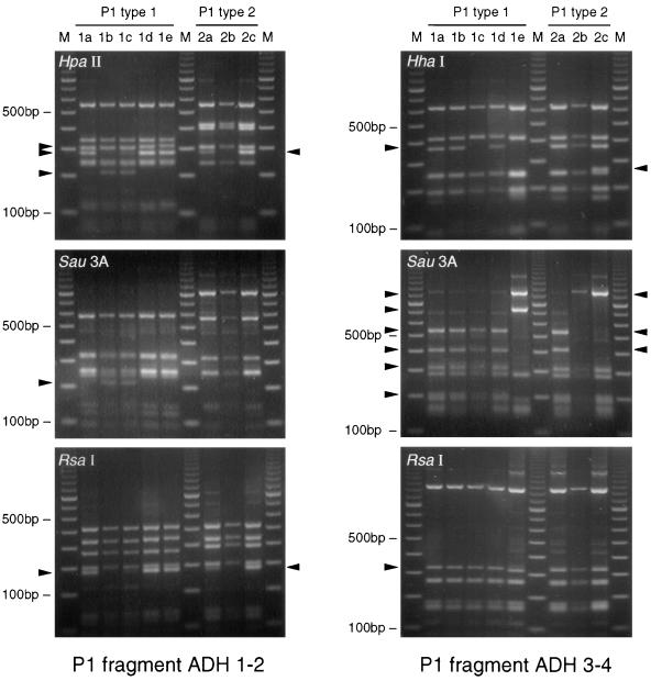
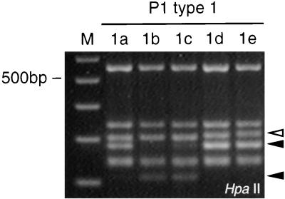
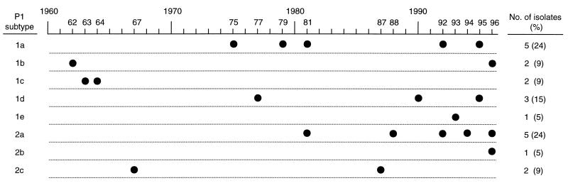
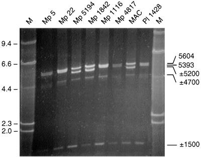
Three methods for genotyping of Mycoplasma pneumoniae clinical isolates were applied to 2 reference strains and 21 clinical isolates. By a modified restriction fragment length polymorphism (RFLP) analysis of PCR products of the M. pneumoniae cytadhesin P1 gene, 5 subtypes were discriminated among 13 P1 type 1 strains and 3 subtypes were discriminated among 8 P1 type 2 strains. Sequence analysis of the 16S-23S rRNA gene spacer region and part of the 23S rRNA gene revealed one nucleotide difference in the intergenic spacer region in 3 of the 21 isolates. In the 23S rRNA gene sequence of the 8 P1 type 2 strains an extra adenosine was present, but it was absent from the 13 P1 type 1 strains. On the basis of M. pneumoniae genome sequence data, primers were designed to amplify large interrepeat fragments by long PCR, and these fragments were subsequently analyzed by RFLP analysis. Only two types, long PCR types 1 and 2, could be discriminated among the M. pneumoniae isolates. All P1 type 1 strains were assigned to long PCR type 1, and all P1 type 2 strains were assigned to long PCR type 2. These data obtained by three independent typing methods thus confirm the existence of two distinct M. pneumoniae genomic groups but expand the possibility of strain typing on the basis of variations within their P1 genes.


Figures

FIG. 1
FIG. 1
PCR-RFLP patterns of two PCR fragments of the P1 gene of M. pneumoniae isolates digested with four endonucleases. Lanes 1a to 1e, M. pneumoniae strains with P1 subtype designation 1a to 1e, respectively, in Table 3; lanes 2a to 2c, M. pneumoniae strains with subtype designation 2a to 2c, respectively, in Table 3; lane M, 100-bp DNA ladder (Promega, Madison, Wis.). The arrowheads on the left side of each panel indicate differences in banding patterns among the P1 type 1 strains, and the arrowheads on the right side of each panel indicate differences in banding patterns among the P1 type 2 strains.
FIG. 2
FIG. 2
Detailed representation of patterns of ADH1-ADH2-generated fragments of P1 type 1 strains after digestion of these fragments with HpaII, illustrating the differentiation into subtypes 1a through 1e. Open arrowhead, band differentiating subtype 1a, 1b, and 1c versus 1d and 1e; solid arrowheads, bands differentiating subtypes 1a, 1d, and 1e from subtypes 1b and 1c.
FIG. 3
FIG. 3
Distribution of the eight P1 subtypes among M. pneumoniae clinical isolates isolated in the period from 1962 through 1996.
FIG. 4
FIG. 4
PCR patterns after EL PCR with primer set Rep2/3-Rep5 of representative M. pneumoniae clinical isolates of the eight P1 subtypes (Table 3). Strains PI 1428, Mp 4817, Mp 1116, Mp 22, and Mp 5 are long PCR type 1 strains. Strains MAC, Mp 1842, and Mp 5194 are long PCR type 2 strains. The sizes of the indicated fragments of 5,604 and 5,393 bp (as indicated on the right) were calculated on the basis of the sequence data for M. pneumoniae (12). The indicated fragments (on the right) of approximately 5,200, 4,700, and 1,500 bp were not expected on the basis of the genome data (12). The approximate molecular sizes of these amplicons were calculated on the basis of their migrations in the gel relative to those of the fragments of the molecular size marker II (lane M; Boehringer) (the numbers on the left are in kilodaltons).

References

    1. Baseman J B, Cole R M, Krause D C, Leith D K. Molecular basis for cytadsorption of Mycoplasma pneumoniae. J Bacteriol. 1982;151:1514–1522. - PMC - PubMed
    1. Bergmans A M, Schellekens J F, van Embden J D, Schouls L M. Predominance of two Bartonella henselae variants among cat-scratch disease patients in The Netherlands. J Clin Microbiol. 1996;34:254–260. - PMC - PubMed
    1. De Wit M Y, Klatser P R. Mycobacterium leprae isolates from different sources have identical sequences of the spacer region between the 16S and 23S ribosomal RNA genes. Microbiology. 1994;140:1983–1987. - PubMed
    1. Don R H, Cox P T, Wainwright B J, Baker K, Mattick J S. ‘Touchdown’ PCR to circumvent spurious priming during gene amplification. Nucleic Acids Res. 1991;19:4008. - PMC - PubMed
    1. Dorigo-Zetsma J W, Zaat S A J, Wertheim-van Dillen P M E, Spanjaard L, Rijntjes J, van Waveren G, Jensen J S, Angulo A F, Dankert J. Comparison of PCR, culture, and serological tests for diagnosis of Mycoplasma pneumoniae respiratory tract infection in children. J Clin Microbiol. 1999;37:14–17. - PMC - PubMed

MeSH terms

LinkOut - more resources