. 2000 Mar 1;523 Pt 2(Pt 2):377-89.
doi: 10.1111/j.1469-7793.2000.t01-2-00377.x.

Functional expression and regulation of the hyperpolarization activated non-selective cation current in embryonic stem cell-derived cardiomyocytes

Affiliations

Functional expression and regulation of the hyperpolarization activated non-selective cation current in embryonic stem cell-derived cardiomyocytes

N Abi-Gerges et al. J Physiol. .

Abstract

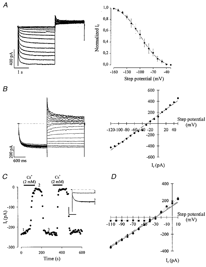
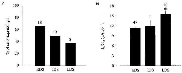
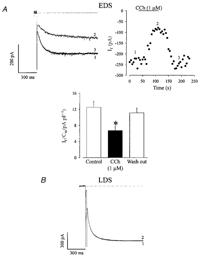
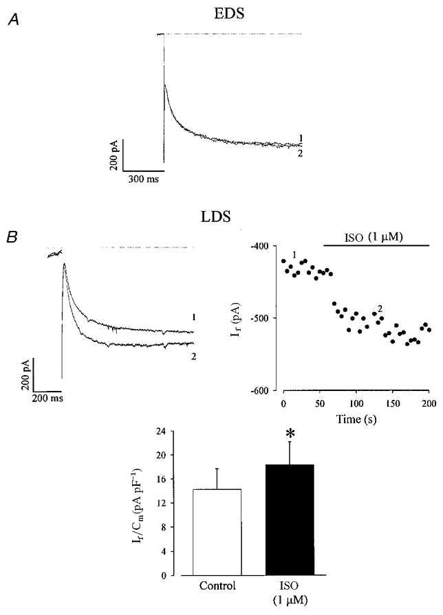
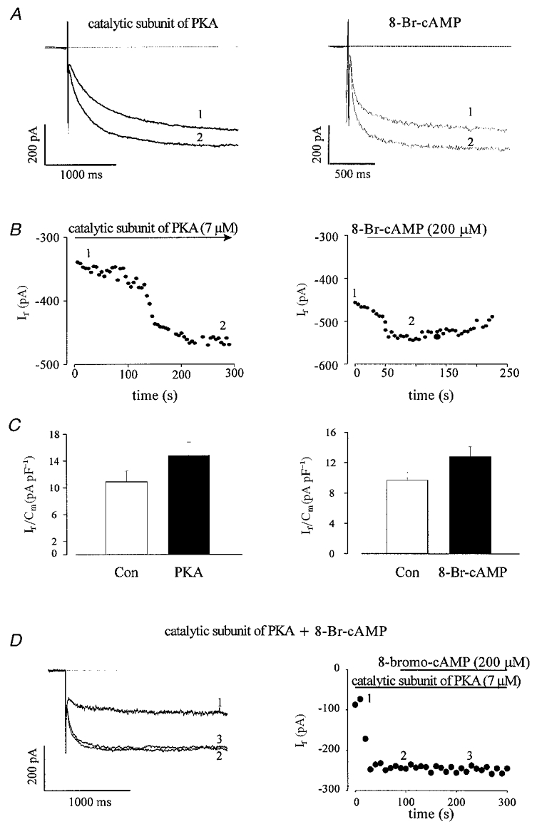
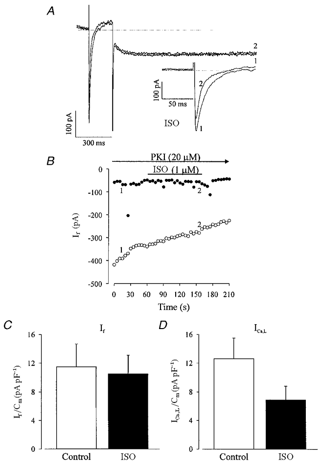
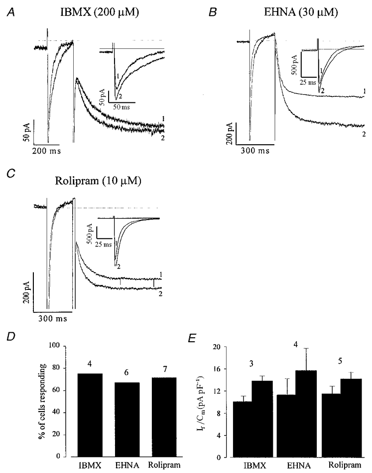
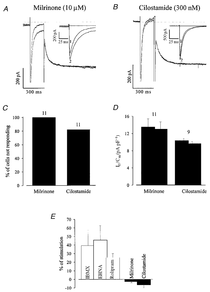
1. The biophysical and pharmacological characteristics of the hyperpolarization activated non- selective cation current (If) were recorded using whole-cell voltage clamp in embryonic stem (ES) cell-derived cardiomyocytes at different stages of development. 2. The cation current was detected in a large percentage (65 %) of early stage (EDS, differentiated for 7 + 3-4 days) cells at a current density of 11.4 +/- 0.6 pA pF-1 (n = 47). In late stage (LDS, differentiated for 7 + 9-12 days) cells the percentage of cells expressing If decreased (45 %), but If densities (15.5 +/- 0.9 pA pF-1, n = 20) were increased. 3. The muscarinic agonist carbachol (CCh, 1 microM) depressed basal If in EDS cells by 45.7 +/- 6.5 %, n = 5) and was without effect in LDS cardiomyocytes (n = 4). The beta-adrenoceptor agonist isoprenaline (ISO, 1 microM) stimulated If in LDS cells by 33 +/- 5.2 % (n = 6) but not in EDS cells (n = 5). 4. Cell infusion with the catalytic subunit of the cAMP-dependent protein kinase (PKA, 7 microM) stimulated If in EDS cells by 37.0 +/- 2.9 %, (n = 4), but subsequent superfusion of 8-bromo-cAMP (200 microM) was without effect. Intracellular perfusion of LDS cardiomyocytes with the highly selective peptide inhibitor of PKA (PKI, 20 microM) completely inhibited the stimulation of the L-type Ca2+ current (ICa,L) as well as of If by ISO (1 microM). 5. Extracellular superfusion with phosphodiesterase (PDE) inhibitors - IBMX, a non-selective antagonist, Erythro-9-(2-hydoxy-3-nonyl)adenine (EHNA), a PDE2 antagonist and rolipram, a PDE4 antagonist - resulted in stimulation of ICa,L and If in EDS cells. By contrast, milrinone and cilostamide, two PDE3 antagonists, stimulated ICa,L, but not If. 6. The present work demonstrates that If is functionally expressed during early cardiomyogenesis. Similar to ICa,L, If is regulated during embryonic development by phosphorylation via PKA. In contrast to ICa,L, If is not regulated by PDE3 suggesting different localization of these ion channels with respect to PDE3.


Figures

Figure 1
Figure 1. Biophysical and pharmacological characterization of If in ES cell-derived cardiomyocytes
A, left panel, IDS cardiomyocyte displaying the steady-state activation characteristics tested by 2 s lasting hyperpolarizing voltage steps from −40 to −160 mV in 10 mV intervals and subsequent 2 s depolarization to +15 mV (holding potential −35 mV, the dotted line indicates the zero current level). The typical slow activation kinetics for If was observed, the threshold potential proved close to −50 mV. The right panel shows the steady-state activation characteristics of 3 representative experiments. Data points were obtained by measuring the peak of the deactivating tail currents, normalized and fitted with a Boltzmann curve yielding a Vh of −98.2 ± 1.1 mV. B, a representative I–V relationship of an EDS cardiomyocyte obtained by applying 2 s lasting prepulses to −115 mV (holding potential −35 mV) and subsequent 2 s lasting voltage steps ranging from −120 to +50 mV in 10 mV intervals. The I–V relationship (right panel) was determined by measuring If 15 ms after applying the depolarizing voltage steps. The linear regression of the data point yielded a reversal potential of −29 mV. C, extracellular application of Cs+ (2 mM) resulted in a complete block of If (holding potential −35 mV, step potential −110 mV) in an EDS cardiomyocyte; this could be reversed by washout (left panel). The numbers in the time course diagram indicate when the current traces displayed in the inset (calibration bars correspond to 300 ms and 200 pA, respectively) were recorded (the current in the presence of Cs+ was taken as reference value for the estimation of If amplitude). The right panel shows the I–V relation of If in absence and presence of extracellular Cs+ (2 mM) in an EDS cardiomyocyte. Cs2+ almost completely blocked If at potentials negative to the reversal potential (the linear regression yielded −30 mV prior to application of Cs+ and −32.5 mV after washout of Cs+) whereas positive to −30 mV no effect of Cs+ on If was observed (•, control; ▪, in the presence of 2 mM CsCl; ▴, after washout of CsCl).
Figure 2
Figure 2. Functional expression of If in ES cell-derived cardiomyocytes at different stages of development
A, the percentage of cells expressing If declined during cardiomyogenesis. B, at later stages of development an increase in the current density (If/Cm, where Cm is membrane capacitance) of If was observed. If was evoked by applying a 2 s hyperpolarizing voltage step from a holding potential of −35 to −110 mV (*statistically significantly different to EDS and IDS at the P < 0.05 level using Student's unpaired t test). Numbers above bars are numbers of replicates.
Figure 3
Figure 3. Carbachol (CCh) strongly depresses the amplitude of basal If in EDS cardiomyocytes
A, CCh depressed basal If in a representative EDS cardiomyocyte. The effect was completely reversed upon washout (left panel). This is also seen in the time course of this experiment (right panel), the numbers indicate the traces displayed in A (the instantaneous current in presence of CCh was taken as reference for the estimation of the If amplitude). The effect of CCh on If in EDS cells (n= 5) is summarized in the histogram. *Statistically significantly different to control at the P < 0.05 level (paired t test). B, in LDS cardiomyocytes CCh did not depress basal If. (Voltage protocol identical to Fig. 2). 1, control; 2, CCh (1 μM).
Figure 4
Figure 4. Isoprenaline (ISO) stimulates If in LDS cardiomyocytes
A, ISO (1 μM) did not stimulate If in EDS cardiomyocytes, 1, control; 2, ISO (1 μM). B, ISO stimulated If in LDS cardiomyocytes (left panel: original recordings; right panel: time course of this experiment, voltage protocol identical to Fig. 2). *Difference in current density between the absence and the presence of ISO was statistically significant (B, histogram, n= 6; P < 0.05, paired t test).
Figure 5
Figure 5. The catalytic subunit of PKA (C-subunit of PKA, 7 μM) and 8-bromo-cAMP (200 μM) stimulate If in EDS cardiomyocytes
A, the C-subunit of PKA (7 μM through pipette, left panel) as well as 8-bromo-cAMP (8-Br-cAMP 200 μM, extracellular application, right panel) stimulated If in EDS cells. The corresponding time courses are shown in B. The effects of C-subunit of PKA (n= 4) and 8-Br-cAMP (n= 5) on If are summarized in the left and right panels of C, respectively. *Statistically significantly different to control (Con) at the P < 0.05 level (paired t test). D shows that 8-Br-cAMP has no additional effect on If after activation of the current by the C-subunit of PKA (7 μM): left panel, current traces (1 control, 2 C-subunit of PKA, 3 C-subunit of PKA + 8-bromo-cAMP); right panel, corresponding time course.
Figure 6
Figure 6. Effect of a selective peptide inhibitor of cAMP-dependent protein kinase A (PKI, 20 μM) on the ISO-stimulated If and L-type Ca2+ current (ICa,L) in LDS cells
A, ISO neither stimulated If nor ICa,L in the presence of PKI (1 PKI, 2 PKI and ISO). The inset in A displays ICa,L at a more extended time scale. Simultaneous recording of ICa,L and If consisted of a depolarizing step from a holding potential of −50 to 0 mV for 200 ms and a subsequent hyperpolarization to −110 mV for 2 s. B, time course of the experiment displayed in A. The If (•) amplitude remained unaltered upon addition of ISO whereas the pronounced decrease in ICa,L (^) was due to run down. A statistical summary of these data demonstrates that neither If (C; n= 6) nor ICa,L (D; n= 4) was stimulated by ISO in the presence of PKI.
Figure 7
Figure 7. The non-selective phosphodiesterase (PDE) inhibitor IBMX as well as PDE2 and PDE4 antagonists stimulate If and ICa,L in EDS cardiomyocytes
A–C, the non-selective antagonist IBMX (200 μM; A), the PDE2 selective antagonist EHNA (30 μM; B) and the PDE4 antagonist rolipram (10 μM; C) stimulate both If and ICa,L in EDS cardiomyocytes. The same voltage protocol as in Fig. 6 was used. The insets display ICa,L at a more extended time scale. Due to the slight delay in the maximal response of If as compared with ICa,L and the fast run down of the latter, the ICa,L traces displayed in B and C were recorded prior to If. D shows that most of the EDS cardiomyocytes tested responded to PDE antagonist application with a stimulation of If and ICa,L. E shows that an increase in If density was also observed under these conditions.
Figure 8
Figure 8. The PDE3 antagonists milrinone and cilostamide stimulate ICa,L but not If in EDS cardiomyocytes
A and B, milrinone and cilostamide stimulated ICa,L (see also inset for a more extended time scale), but If remains unaltered (voltage protocol identical to Fig. 6). C, in the large majority of cells If is not stimulated upon PDE3 antagonist application. D, the current density of the non-responding cells decreased slightly in the presence of PDE inhibitors indicating a small run down. E, a quantitative comparison of the percentage of If density stimulation demonstrates that If was stimulated by PDE2 and 4 antagonists, but not by PDE3 antagonists.

References

    1. An RH, Davies MP, Doevendans PA, Kubalak SW, Bangalore R, Chien KR, Kass RS. Developmental changes in beta-adrenergic modulation of L-type Ca2+ channels in embryonic mouse heart. Circulation Research. 1996;78:371–378. - PubMed
    1. An RH, Heath BM, Koch W, Lefkowitz RJ, Kass RS. β2-Adrenergic receptor overexpression in the developing mouse heart: evidence for targeted modulation of ion channels. The Journal of Physiology. 1999;516:19–30. - PMC - PubMed
    1. Bois P, Lenfant J. Isolated cells of the frog sinus venosus: properties of the inward current activated during hyperpolarization. Pflügers Archiv. 1990;416:339–346. - PubMed
    1. Bois P, Renaudon B, Baruscotti M, Lenfant J, Difrancesco D. Activation of f-channels by cAMP analogues in macropatches from rabbit sino-atrial node myocytes. The Journal of Physiology. 1997;501:565–571. - PMC - PubMed
    1. Callewaert G, Carmeliet E, Vereecke J. Single cardiac Purkinje cells: general electrophysiology and voltage-clamp analysis of the pace-maker current. The Journal of Physiology. 1984;349:643–661. - PMC - PubMed

Publication types

MeSH terms

Substances

LinkOut - more resources