

Blockade of endogenous interleukin 12 results in suppression of murine streptococcal cell wall arthritis by enhancement of interleukin 10 and interleukin 1Ra
- PMID: 10700428
- PMCID: PMC1753084
- DOI: 10.1136/ard.59.3.196
Blockade of endogenous interleukin 12 results in suppression of murine streptococcal cell wall arthritis by enhancement of interleukin 10 and interleukin 1Ra
Abstract
Objective: The goal of this study was to investigate the role of endogenous interleukin 12 (IL12) in acute murine streptococcal cell wall (SCW) arthritis.
Methods: C57black/6 mice were injected intraperitoneally with rat anti-murine IL12 (C17.8), shortly before induction of arthritis by intra-articular injection of 25 microg SCW fragments into the right knee joint. Joint swelling and chondrocyte synthetic function was analysed several days after induction of SCW arthritis. Local cytokine profile was determined, protein by using ELISA and mRNA by RT-PCR technology. To confirm the findings at later time points, tissue chamber model of inflammation was used. Histology was performed to examine cell influx and cartilage damage.
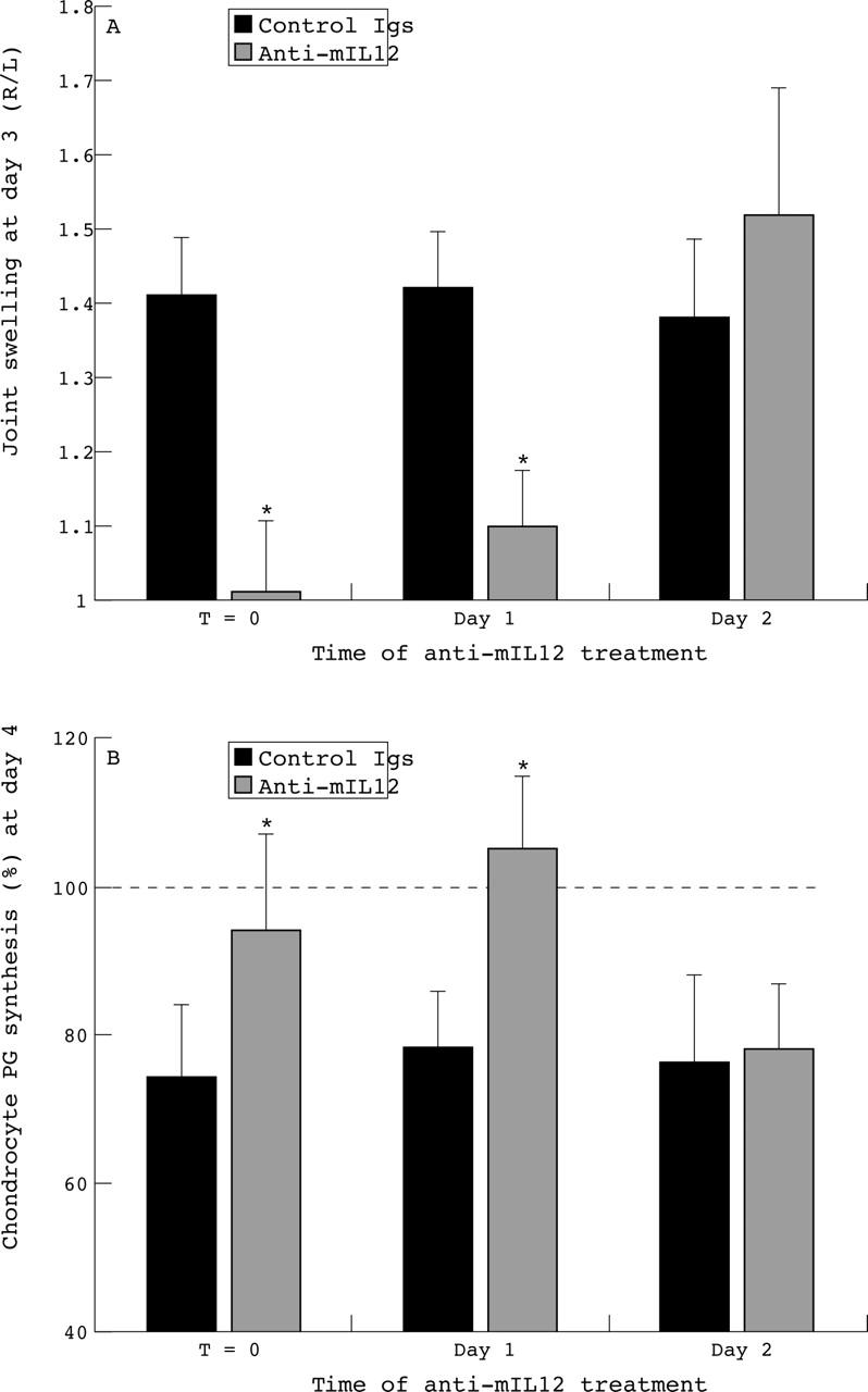
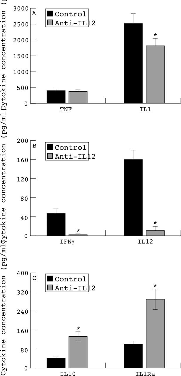
Results: Suppression of joint swelling was noted at days 2 and 4, whereas no suppressive effect of anti-IL12 was found at day 1. Severe inhibition of chondrocyte proteoglycan synthesis was seen at day 1 in both arthritic control and anti-IL12 treated mice. However, chondrocyte function was restored at day 4 of arthritis in the anti-IL12 injected animals, but not in the arthritic controls. Moreover, cell influx in synovial tissue and joint cavity was reduced by anti-IL12 treatment. Neutralisation of IL12 reduced the local levels of IL1beta, IL12 and interferon gamma, when examined shortly after induction of SCW arthritis, whereas tumour necrosis factor alpha levels were not affected. In contrast, IL10 and IL1Ra protein and mRNA levels were strongly up regulated in synovial tissues after IL12 blockade. Enhancement of IL10 and IL1Ra by anti-IL12 was confirmed in a tissue chamber model with SCW induced inflammation.
Conclusions: This study indicates that IL12 is a pro-inflammatory cytokine during onset of acute SCW arthritis. Balances of proinflammatory and anti-inflammatory cytokines were strongly improved by anti-IL12 treatment.
Figures


Similar articles
-
An IFN-gamma-independent proinflammatory role of IL-18 in murine streptococcal cell wall arthritis.J Immunol. 2000 Dec 1;165(11):6553-8. doi: 10.4049/jimmunol.165.11.6553. J Immunol. 2000. PMID: 11086098
-
Different roles of tumour necrosis factor alpha and interleukin 1 in murine streptococcal cell wall arthritis.Cytokine. 1998 Sep;10(9):690-702. doi: 10.1006/cyto.1998.0372. Cytokine. 1998. PMID: 9770330
-
Local interleukin-12 gene transfer promotes conversion of an acute arthritis to a chronic destructive arthritis.Arthritis Rheum. 2002 May;46(5):1379-89. doi: 10.1002/art.10233. Arthritis Rheum. 2002. PMID: 12115246
-
Gene therapy for rheumatoid arthritis. Lessons from animal models, including studies on interleukin-4, interleukin-10, and interleukin-1 receptor antagonist as potential disease modulators.Rheum Dis Clin North Am. 2002 Feb;28(1):127-49. doi: 10.1016/s0889-857x(03)00073-5. Rheum Dis Clin North Am. 2002. PMID: 11840694 Review.
-
Arthritic and non-arthritic synovial fluids modulate IL10 and IL1RA gene expression in differentially activated primary human monocytes.Osteoarthritis Cartilage. 2015 Nov;23(11):1853-7. doi: 10.1016/j.joca.2015.06.003. Osteoarthritis Cartilage. 2015. PMID: 26521731 Review.
Cited by
-
Rheumatoid arthritis vaccine therapies: perspectives and lessons from therapeutic ligand epitope antigen presentation system vaccines for models of rheumatoid arthritis.Expert Rev Vaccines. 2015 Jun;14(6):891-908. doi: 10.1586/14760584.2015.1026330. Epub 2015 Mar 18. Expert Rev Vaccines. 2015. PMID: 25787143 Free PMC article. Review.
-
GM-CSF neutralisation suppresses inflammation and protects cartilage in acute streptococcal cell wall arthritis of mice.Ann Rheum Dis. 2007 Apr;66(4):452-7. doi: 10.1136/ard.2006.057182. Epub 2006 Oct 4. Ann Rheum Dis. 2007. PMID: 17020908 Free PMC article.
-
Gut permeability and osteoarthritis, towards a mechanistic understanding of the pathogenesis: a systematic review.Ann Med. 2021 Dec;53(1):2380-2390. doi: 10.1080/07853890.2021.2014557. Ann Med. 2021. PMID: 34933614 Free PMC article.
-
Treating experimental arthritis with the innate immune inhibitor interleukin-37 reduces joint and systemic inflammation.Rheumatology (Oxford). 2016 Dec;55(12):2220-2229. doi: 10.1093/rheumatology/kew325. Epub 2016 Aug 26. Rheumatology (Oxford). 2016. PMID: 27567100 Free PMC article.
-
NK cells help to induce CD8(+)-T-cell immunity against Toxoplasma gondii in the absence of CD4(+) T cells.Infect Immun. 2005 Aug;73(8):4913-21. doi: 10.1128/IAI.73.8.4913-4921.2005. Infect Immun. 2005. PMID: 16041005 Free PMC article.
References
Publication types
MeSH terms
Substances
LinkOut - more resources
Full Text Sources
Other Literature Sources
Medical
