Comparative Study
. 2000 Mar;156(3):857-64.
doi: 10.1016/S0002-9440(10)64954-1.

A novel association between the human heat shock transcription factor 1 (HSF1) and prostate adenocarcinoma

Affiliations
Comparative Study

A novel association between the human heat shock transcription factor 1 (HSF1) and prostate adenocarcinoma

A T Hoang et al. Am J Pathol. 2000 Mar.

Abstract

A search for differentially expressed genes in a pair of nonmetastatic (PC-3) versus metastatic variant (PC-3M) human prostate carcinoma cell lines led to identification of the human heat shock factor (HSF1) as an overexpressed gene product in PC-3M cells. Analysis of primary prostate cancer specimens indicated that HSF1 is generally up-regulated in most of the malignant prostate epithelial cells relative to the normal prostate cells. Among the known effectors of HSF1 action, constitutive levels of HSP70 and HSP90 are not significantly altered by the naturally elevated expression of HSF1 as in PC-3M cells or by transduced overexpression of HSF1 in PC-3 cells. The basal levels of HSP27 in both cases are, however, consistently increased by two- to threefold. With respect to response to heat shock, high basal concentration of HSP90 is not further enhanced in these cells, and HSP70 is up-regulated irrespective of HSF1 level. Heat shock, however, causes an increase in HSP27 when HSF1 is up-regulated, except when the expression of HSF1 is already too high. These results document for the first time that HSF1 is overexpressed in human prostate cancer cells, at least one consequence of which in the prostate cancer cell lines tested is stimulation of both basal and stress-induced expression of HSP27, an important factor in cell growth, differentiation, or apoptosis.


Figures

Figure 1.
Figure 1.
Northern blot showing differential expression of HSF1 in PC-3 and PC-3M cell lines. Total RNA (10 μg) from PC-3 (lane 1) or PC-3M (lane 2) cells was analyzed using the 250-bp HSF1 fragment as probe. The same blot was subsequently hybridized to β-actin probe as an RNA loading control.
Figure 2.
Figure 2.
Western blots illustrating the expression of HSF1 protein in prostate cell lines and tissues. A: Cells used: 1, immortalized prostate epithelial (MLC); 2, LNCaP; 3, LAPC-4; 4, PC-3; 5, PC-3M; 6, DU-145. B: Prostate tissue sections: 1, normal (S95–1066); 2, normal (S15113); 3, normal (S15121); 4, tumor S96–158; 5, tumor S15050; and 6, tumor S15284. The lower part of A shows α-actin as loading control; the lower part of B depicts Coomassie Blue staining of the gels.
Figure 3.
Figure 3.
Immunohistochemical staining of HSF1 protein in PC-3 and PC-3M cells. Cytospins of PC-3 and PC-3M cells were stained with both hematoxylin and anti-HSF1 antibodies.
Figure 4.
Figure 4.
Illustration of HSF1 immunostaining in normal and carcinomatous prostate tissue sections. Formalin-fixed frozen sections were stained with both hematoxylin and anti-HSF1 antibodies. A: Normal section (S95–1068) B: Tumor section (S15–2484). C: Normal section from the specimen P735. D: Tumor section from the same specimen, P735.
Figure 5.
Figure 5.
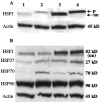
Western blots to determine the levels of expression of heat shock proteins in PC-3 and PC-3M cells before and after heat shock treatment. After heat shock, the cells were either analyzed immediately (A) or after 4 hours’ recovery time (B). Lane 1: PC-3; lane 2: PC-3 heat-treated; lane 3: PC-3M; lane 4: PC-3M heat-treated. The blot was first examined for HSF1, and then stripped and reused sequentially for HSP27, HSP70, HSP90, and α-actin. p, phosphorylated; un, unphosphorylated species of HSF1.
Figure 6.
Figure 6.
Effect of HSF1 overexpression in PC-3 cells with respect to heat shock protein levels. The results are illustrated from a PC-3 vector clone (PC-3V3) and a PC-3 HSF1-overexpressed clone (PC-3H16/3). Cells were heated at 45°C for 1 hour and cell lysates were harvested immediately (A) or after 4 hours’ recovery (B). Antibodies for HSF1, HSP27, HSP70, HSP90, and α-actin were used as indicated. Lane 1: PC-3V3 control; lane 2: PC-3V3 heat shock; lane 3: PC-3H16/3 control; lane 4: PC-3H16/3 heat shock. p, phosphorylated form of HSF1; un, unphosphorylated form of HSF1.
Figure 7.
Figure 7.
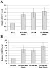
Quantitation of HSP27 levels by densitometry normalized with respect to α-actin loading. HSP27 level in PC-3 (A) or PC-3V3 (B) was set as 1. Error bars represent SD from three independent experiments in each case.

Similar articles

Cited by

References

    1. Rabindran SK, Giorgi J, Clos J, Wu C: Molecular cloning and expression of a human heat shock factor, HSF1. Proc Natl Acad Sci USA 1991, 88:6906-6910 - PMC - PubMed
    1. Sarge KD, Zimarino V, Holm K, Wu C, Morimoto RI: Cloning and characterization of two mouse heat shock factors with distinct inducible and constitutive DNA-binding ability. Genes Dev 1991, 5:1902-1911 - PubMed
    1. Schuetz TJ, Gallo GJ, Sheldon L, Tempst P, Kingston RE: Isolation of a cDNA for HSF2: evidence for two heat shock factor genes in humans. Proc Natl Acad Sci USA 1991, 88:6911-6915 - PMC - PubMed
    1. Wu C: Heat shock transcription factors: structure and regulation. Ann Rev Cell Dev Biol 1995, 11:441-469 - PubMed
    1. Nakai A, Tanabe M, Kawazoe Y, Inazawa J, Morimoto RI, Nagata K: HSF4, a new member of the human heat shock factor family which lacks properties of a transcriptional activator. Mol Cell Biol 1997, 17:469-481 - PMC - PubMed

Publication types

MeSH terms