Comparative Study
. 2000 Mar;156(3):951-64.
doi: 10.1016/S0002-9440(10)64963-2.

Expression of human apolipoprotein E4 in neurons causes hyperphosphorylation of protein tau in the brains of transgenic mice

Affiliations
Comparative Study

Expression of human apolipoprotein E4 in neurons causes hyperphosphorylation of protein tau in the brains of transgenic mice

I Tesseur et al. Am J Pathol. 2000 Mar.

Abstract

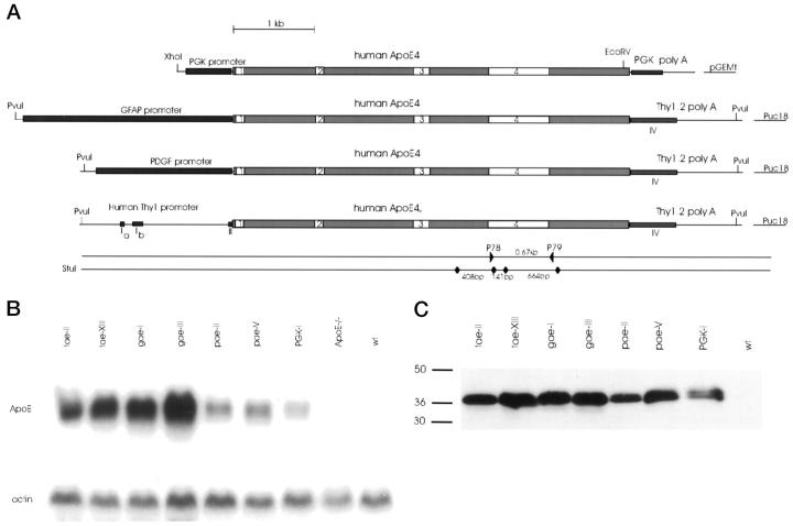
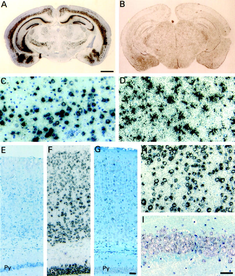
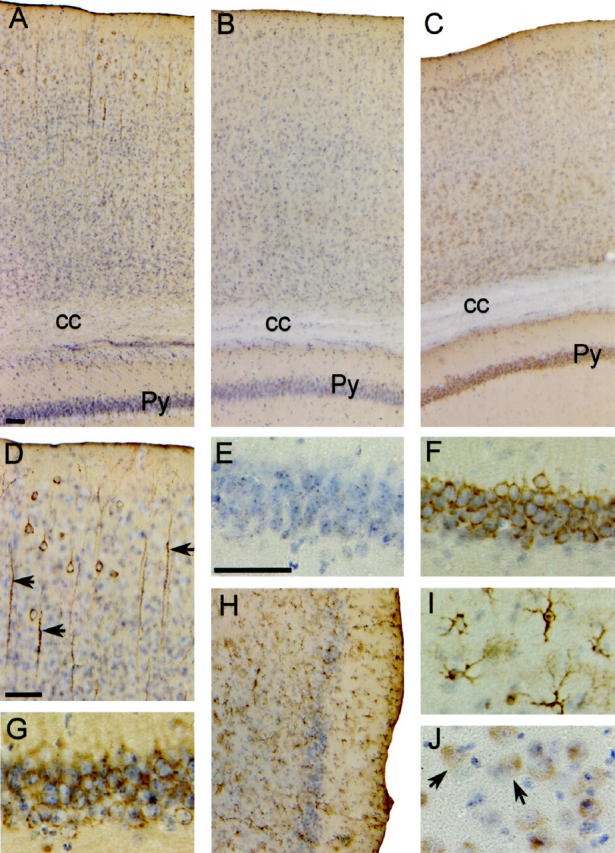
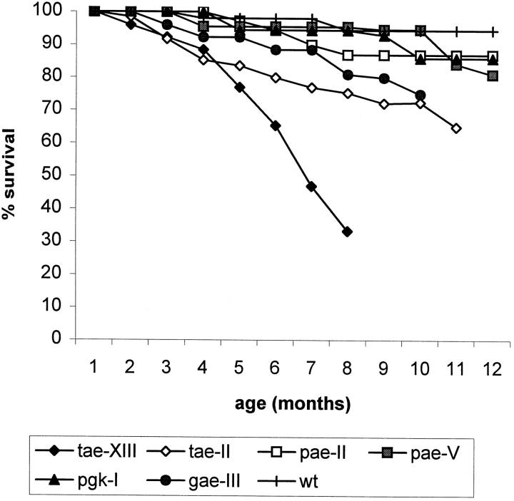
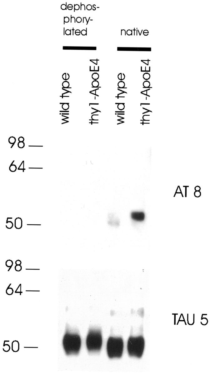
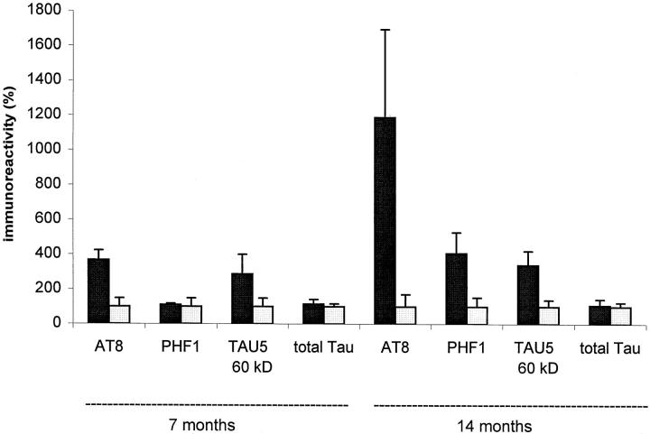
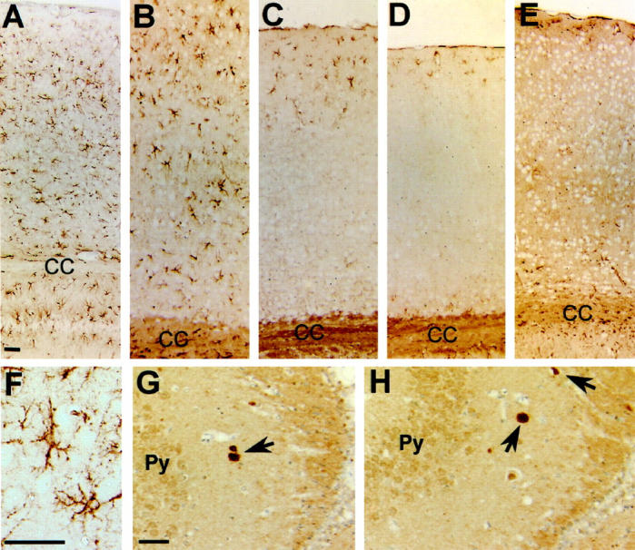
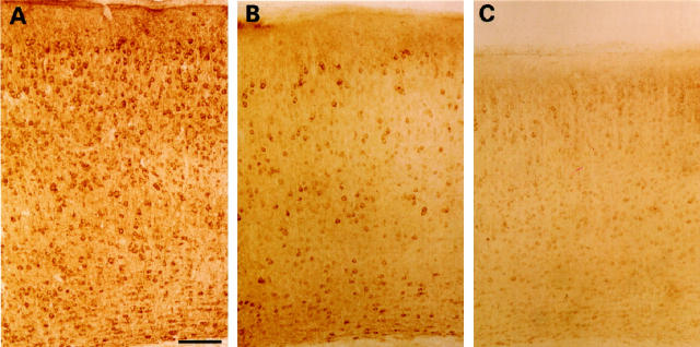
Epidemiological studies have established that the epsilon 4 allele of the ApoE gene (ApoE4) constitutes an important risk factor for Alzheimer's disease and might influence the outcome of central nervous system injury. The mechanism by which ApoE4 contributes to the development of neurodegeneration remains unknown. To test one hypothesis or mode of action of ApoE, we generated transgenic mice that overexpressed human ApoE4 in different cell types in the brain, using four distinct gene promoter constructs. Many transgenic mice expressing ApoE4 in neurons developed motor problems accompanied by muscle wasting, loss of body weight, and premature death. Overexpression of human ApoE4 in neurons resulted in hyperphosphorylation of the microtubule-associated protein tau. In three independent transgenic lines from two different promoter constructs, increased phosphorylation of protein tau was correlated with ApoE4 expression levels. Hyperphosphorylation of protein tau increased with age. In the hippocampus, astrogliosis and ubiquitin-positive inclusions were demonstrated. These findings demonstrate that expression of ApoE in neurons results in hyperphosphorylation of protein tau and suggests a role for ApoE in neuronal cytoskeletal stability and metabolism.


Figures

Figure 1.
Figure 1.
Generation and characterization of different ApoE4 transgenic mouse strains. A: Schematic representation of minigene constructs synthesized as described in the text. On the lower lines are indicated the StuI restriction site and size of restriction fragments used in genotyping by Southern blotting. The polymerase chain reaction primers (P78 and P79) used for routine genotyping are also indicated. B: Northern blot analysis of representative mice from the seven different ApoE4 transgenic strains. Total RNA (10 μg) was sequentially analyzed for expression of human ApoE mRNA (human-specific probe) and for actin mRNA. RNA from an ApoE knockout mouse and a wild-type mouse (wt) is included. C: Western blot of proteins extracted from the brains of the same mice as used in panel B, except for the ApoE knockout mice. A human-specific anti-human ApoE antibody was used (see Materials and Methods).
Figure 2.
Figure 2.
In situ hybridization of brains of ApoE4 transgenic and Wt mice. Sections were probed with the antisense human-specific ApoE probe and counterstained with toluidine blue. Coronal brain sections of Thy1-ApoE4 (A), and GFAP-ApoE4 (B) mice. Scale bar, 1 mm. Bar indicated in A also applies to B. C: Detail of the cortex of Thy1-ApoE4 (C) and GFAP-ApoE4 (D) mice. Parietal associative cortex of Wt (E), PDGF-ApoE4 (F), and PGK-ApoE4 (G) mice. Detail of the cortex of PDGF-ApoE4 (H) and of the pyramidal cells of the CA1 region of the hippocampus of PGK-ApoE4 (I) mice. Scale bar, 50 μm. Bar indicated in G also applies to E and F. Bar indicated in I also applies to C, D, and H. Py, pyramidal cell layer of the hippocampus.
Figure 3.
Figure 3.
Immunohistochemistry for human ApoE4 in brains of transgenic and Wt mice. Sections were counterstained with hematoxylin. A–C: Cortical region of Thy1-ApoE4, Wt, and PDGF-ApoE4 mice, respectively. D: Detail of dendrites in cortex of a Thy1-ApoE4 mouse. E–G: Details of the pyramidal cells in the CA1 region of the hippocampus in Wt , PDGF-ApoE4, and Thy1-ApoE4 transgenic mice, respectively. H and I: Astrocytes in the entorhinal cortex of a GFAP-ApoE4 mouse. J: Astrocytes of neuronal cell bodies in the neocortex of a PGK-ApoE4 mouse. Scale bars, 50 μm. Scale bar indicated in A also applies to B, C, and H. Scale bar indicated in E also applies to G, F, I, and J. Py, pyramidal cells of the hippocampus; cc, corpus callosum.
Figure 4.
Figure 4.
Survival of ApoE4 transgenic mice: tae-XIII, n = 15; tae-II, n = 43; pae-II, n = 31; pae-V, n = 19; pgk-I, n = 14; gae-III, n = 21; wt, n = 30.
Figure 5.
Figure 5.
Comparison of protein tau hyperphosphorylation between Thy1-ApoE4 (line tae-XIII) and wild-type (wt) mice at ages 3 to 18 months. Western blots shown are representative examples. Four independent tau-specific monoclonal antibodies (AT8, AT180, PHF1, and TAU5) were used. Goat-anti-mouse peroxidase was used as secondary antibody.
Figure 6.
Figure 6.
Comparison of protein tau hyperphosphorylation between 2-year-old PDGF-ApoE4 (pae-II), GFAP-ApoE4 (gae-I), and wild-type (wt) mice. Goat-anti-mouse peroxidase was used as secondary antibody.
Figure 7.
Figure 7.
Dephosphorylated protein extract of a Thy1-ApoE4 and wild-type mouse with alkaline phosphatase, compared with untreated (native) samples. AT8 and TAU5 monoclonal antibodies were used to detect bands.
Figure 8.
Figure 8.
Quantitative comparison of the levels of hyperphosphorylated protein tau immunoreactivities of 7- and 14-month-old Thy1-ApoE4 transgenic and age-matched control mice. Data are means ± SEMs (bars) of wild type (□, n = 3) and Thy1-ApoE4 transgenic (▪, n = 3) mice. Data were obtained by densitometric analysis of immunoblots and normalized.
Figure 9.
Figure 9.
A–E: GFAP staining of the parietal associative cortex of Thy1-ApoE4 (A), PDGF-ApoE4 (B), PGK-ApoE4 (C), GFAP-ApoE4 (D), and Wt (E) mice. F: Detail of the hippocampus of a Thy1-ApoE4 mouse showing reactive astrogliosis. G and H: Ubiquitin positive inclusions (arrows) can be seen in the CA3 region of the hippocampus of Thy1-ApoE4 (G) and PDGF-ApoE4 (H) mice. Scale bar, 50 μm. Scale bar indicated in A also applies to B, C, D, and E. Scale bar indicated in G also applies to H. Py, pyramidal cells of the hippocampus; CC, corpus callosum.
Figure 10.
Figure 10.
A: Staining with the polyclonal anti-tau antibody B19 shows a widespread somatodendritic localization in the parietal associative cortex of a Thy1-ApoE4 transgenic mouse 18 months old. B: Staining with the monoclonal phosphorylation-dependent antibody AT8 recording a slightly more intense somatodendritic staining in the parietal associative cortex of the Thy1-ApoE4 mice18 months old, compared with aged-matched wild-type mice (C). Scale bar, 100 μm.

References

    1. Corder EH, Saunders AM, Strittmatter WJ, Schmechel DE, Gaskell PC, Small GW, Roses AD, Haines JL, Pericak-Vance MA: Gene dose of apolipoprotein E type 4 allele and the risk of Alzheimer’s disease in late onset families. Science 1993, 261:921-923 - PubMed
    1. Strittmatter WJ, Saunders AM, Goedert M, Weisgraber KH, Dong LM, Jakes R, Huang DY, Pericak-Vance M, Schmechel D, Roses AD: Isoform-specific interactions of apolipoprotein E with microtubule-associated protein tau: implications for Alzheimer disease. Proc Natl Acad Sci USA 1994, 91:11183-11186 - PMC - PubMed
    1. Olichney JM, Hansen LA, Galasko D, Saitoh T, Hofstetter CR, Katzman R, Thal LJ: The apolipoprotein E epsilon 4 allele is associated with increased neuritic plaques and cerebral amyloid angiopathy in Alzheimer’s disease and Lewy body variant. Neurology 1996, 47:190-196 - PubMed
    1. Nagy Z, Esiri MM, Jobst KA, Johnston C, Litchfield S, Sim E, Smith AD: Influence of the apolipoprotein E genotype on amyloid deposition and neurofibrillary tangle formation in Alzheimer’s disease. Neuroscience 1995, 69:757-761 - PubMed
    1. Gearing M, Mori H, Mirra SS: A beta-peptide length and apolipoprotein E genotype in Alzheimer’s disease. Ann Neurol 1996, 39:395-399 - PubMed

Publication types

MeSH terms

LinkOut - more resources