

Prevention of hepatic apoptosis and embryonic lethality in RelA/TNFR-1 double knockout mice
- PMID: 10702415
- PMCID: PMC1876833
- DOI: 10.1016/S0002-9440(10)64967-X
Prevention of hepatic apoptosis and embryonic lethality in RelA/TNFR-1 double knockout mice
Abstract
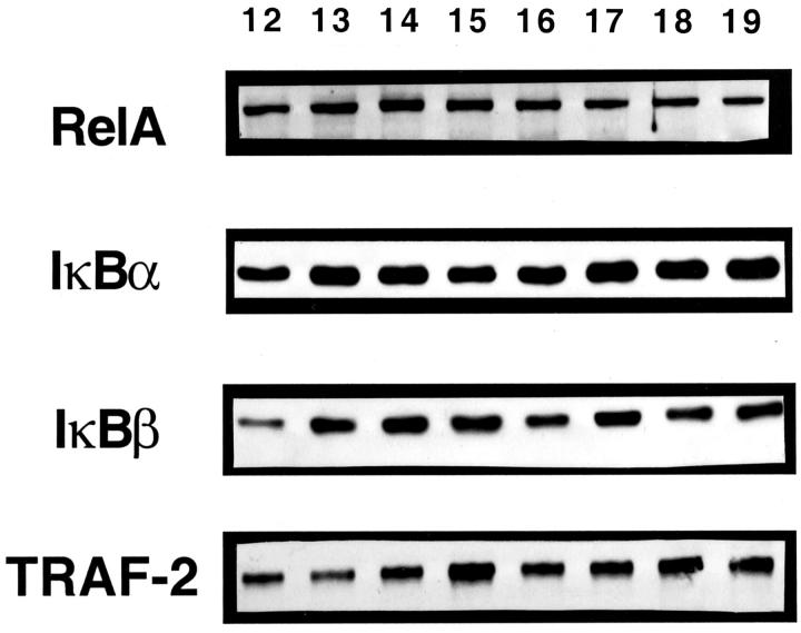
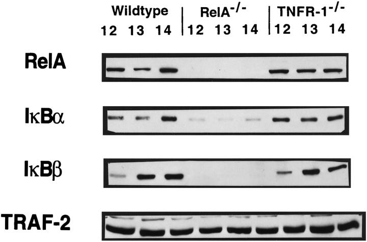
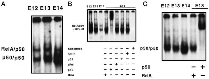
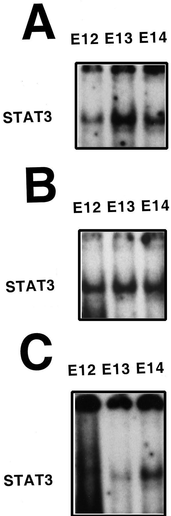
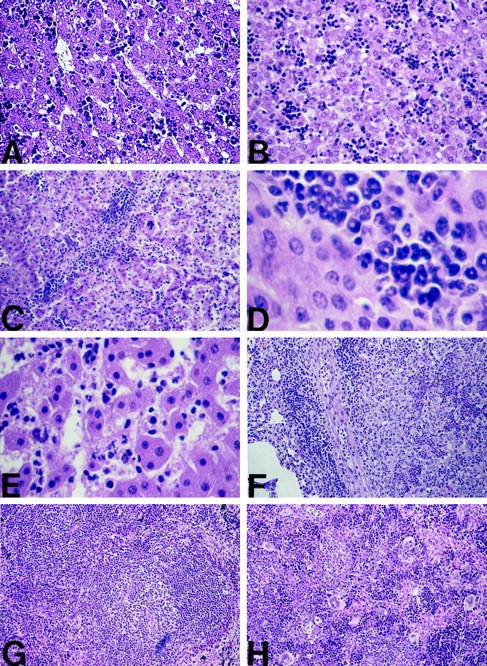
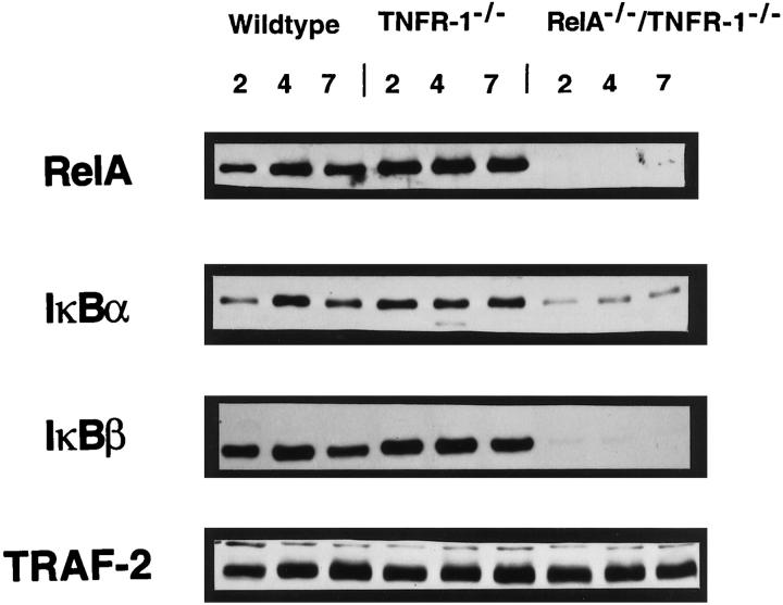
Mice deficient in the nuclear factor kappaB (NF-kappaB)-transactivating gene RelA (p65) die at embryonic days 14-15 with massive liver apoptosis. In the adult liver, activation of the NF-kappaB heterodimer RelA/p50 can cause hepatocyte proliferation, apoptosis, or the induction of acute-phase response genes. We examined, during wild-type fetal liver development, the expression of the Rel family member proteins, as well as other proteins known to be important for NF-kappaB activation. We found these proteins and active NF-kappaB complexes in the developing liver from at least 2 days before the onset of lethality observed in RelA knockouts. This suggests that the timing of NF-kappaB activation is not related to the timing of lethality. We therefore hypothesized that, in the absence of RelA, embryos were sensitized to tumor necrosis factor (TNF) receptor 1 (TNFR-1)-mediated apoptosis. Thus, we generated mice that were deficient in both RelA and TNFR-1 to determine whether apoptotic signaling through TNFR-1 was responsible for the lethal phenotype. RelA/TNFR-1 double knockout mice survived embryonic development and were born with normal livers without evidence of increased hepatocyte apoptosis. These animals became runted shortly after birth and survived an average of 10 days, dying from acute hepatitis with an extensive hepatic infiltration of immature neutrophils. We conclude that neither RelA nor TNFR-1 is required for liver development and that RelA protects the embryonic liver from TNFR-1-mediated apoptotic signals. However, the absence of both TNFR-1 signaling and RelA activity in newborn mice makes these animals susceptible to endogenous hepatic infection.
Figures
Similar articles
-
Analysis of liver regeneration in mice lacking type 1 or type 2 tumor necrosis factor receptor: requirement for type 1 but not type 2 receptor.Hepatology. 1998 Oct;28(4):959-70. doi: 10.1002/hep.510280410. Hepatology. 1998. PMID: 9755232
-
Genetic inactivation of RelA/p65 sensitizes adult mouse hepatocytes to TNF-induced apoptosis in vivo and in vitro.Gastroenterology. 2007 Jun;132(7):2489-503. doi: 10.1053/j.gastro.2007.03.033. Epub 2007 Mar 21. Gastroenterology. 2007. PMID: 17570221
-
SEK1/MKK4-mediated SAPK/JNK signaling participates in embryonic hepatoblast proliferation via a pathway different from NF-kappaB-induced anti-apoptosis.Dev Biol. 2002 Oct 15;250(2):332-47. Dev Biol. 2002. PMID: 12376107
-
To die or not to die: the function of the transcription factor NF-kappaB in embryos exposed to stress.Am J Reprod Immunol. 2004 Feb;51(2):138-43. doi: 10.1046/j.8755-8920.2003.00134.x. Am J Reprod Immunol. 2004. PMID: 14748840 Review.
-
Crosstalk via the NF-kappaB signaling system.Cytokine Growth Factor Rev. 2008 Jun-Aug;19(3-4):187-97. doi: 10.1016/j.cytogfr.2008.04.005. Epub 2008 Jun 2. Cytokine Growth Factor Rev. 2008. PMID: 18515173 Free PMC article. Review.
Cited by
-
NF-kappaB RelA opposes epidermal proliferation driven by TNFR1 and JNK.Genes Dev. 2004 Jan 1;18(1):17-22. doi: 10.1101/gad.1160904. Genes Dev. 2004. PMID: 14724177 Free PMC article.
-
NF-κB RelA Is Required for Hepatoprotection during Pneumonia and Sepsis.Infect Immun. 2019 Jul 23;87(8):e00132-19. doi: 10.1128/IAI.00132-19. Print 2019 Aug. Infect Immun. 2019. PMID: 31160364 Free PMC article.
-
New insights into the role of nuclear factor-kappaB in cell growth regulation.Am J Pathol. 2001 Aug;159(2):387-97. doi: 10.1016/s0002-9440(10)61708-7. Am J Pathol. 2001. PMID: 11485895 Free PMC article. Review.
-
Targeting IKKβ Activity to Limit Sterile Inflammation in Acetaminophen-Induced Hepatotoxicity in Mice.Pharmaceutics. 2023 Feb 20;15(2):710. doi: 10.3390/pharmaceutics15020710. Pharmaceutics. 2023. PMID: 36840032 Free PMC article.
-
TNFR1 determines progression of chronic liver injury in the IKKγ/Nemo genetic model.Cell Death Differ. 2013 Nov;20(11):1580-92. doi: 10.1038/cdd.2013.112. Epub 2013 Aug 9. Cell Death Differ. 2013. PMID: 23933814 Free PMC article.
References
-
- Wulczyn FG, Krappmann D, Scheidereit C: The NF-κB/Rel, and IκB gene families: mediators of immune response and inflammation. J Mol Med 1996, 74:749-769 - PubMed
-
- Sonenshein GE: Rel/NF-κB transcription factors, and the control of apoptosis. Sem Cancer Biol 1997, 8:113-119 - PubMed
-
- Beg AA, Sha WC, Bronson RT, Ghosh S, Baltimore D: Embryonic lethality and liver degeneration in mice lacking the RelA component of NF-κB. Nature 1995, 376:167-170 - PubMed
Publication types
MeSH terms
Substances
Grants and funding
LinkOut - more resources
Full Text Sources
Other Literature Sources
Molecular Biology Databases
Research Materials
