. 2000 Mar;156(3):1025-32.
doi: 10.1016/S0002-9440(10)64970-X.

Bax is increased in the retina of diabetic subjects and is associated with pericyte apoptosis in vivo and in vitro

Affiliations

Bax is increased in the retina of diabetic subjects and is associated with pericyte apoptosis in vivo and in vitro

F Podestà et al. Am J Pathol. 2000 Mar.

Abstract

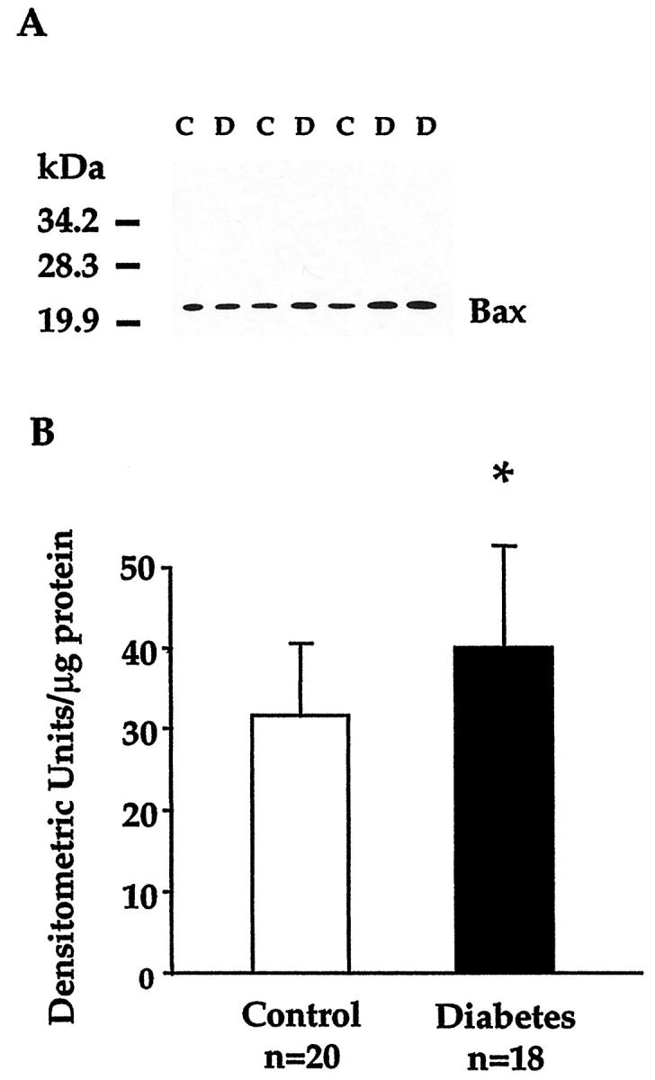
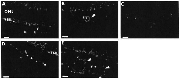
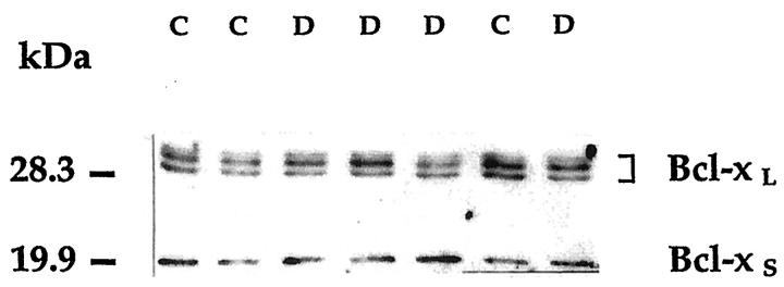
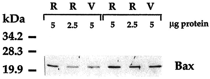
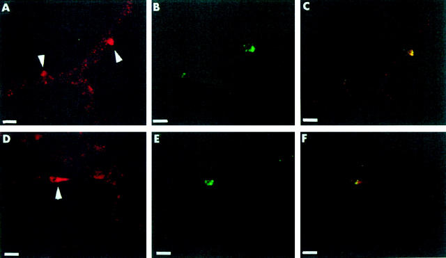
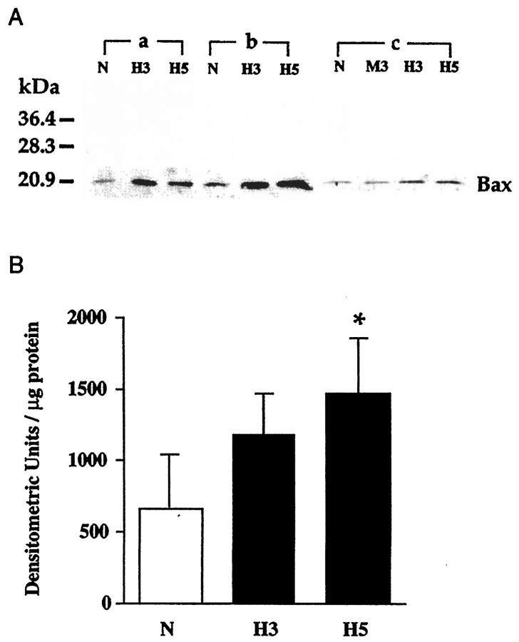
Diabetes of even short duration accelerates the death of capillary cells and neurons in the inner retina by a process consistent with apoptosis. We examined whether the process is accompanied by changes in the expression of endogenous regulators of apoptosis. In postmortem retinas of 18 diabetic donors (age 67 +/- 6 years, diabetes duration 9 +/- 4 years) the levels of pro-apoptotic Bax were slightly, but significantly, increased when compared with levels in 20 age-matched nondiabetic donors (P = 0.04). In both groups, Bax localized to vascular and neural cells of the inner retina. Neither pro-apoptotic Bcl-X(S), nor pro-survival Bcl-X(L) appeared affected by diabetes. The levels of these molecules could not be accurately quantitated in lysates of retinal vessels because of variable degrees of glial contamination. However, studies in situ showed in several pericytes, the outer cells of retinal capillaries, intense Bax staining often in conjunction with DNA fragmentation. Bovine retinal pericytes exposed in vitro to high glucose levels for 5 weeks showed elevated levels of Bax (P = 0.03) and increased frequency of annexin V binding, indicative of early apoptosis. Hence, human diabetes selectively alters the expression of Bax in the retina and retinal vascular pericytes at the same time as it causes increased rates of apoptosis. The identical program induced by high glucose in vitro implicates hyperglycemia as a causative factor in vivo, and provides a model for establishing the role of Bax in the accelerated death of retinal cells induced by diabetes.


Figures

Figure 1.
Figure 1.
Immunoblot analysis of Bax in the human retina. A: Blot representative of the signals obtained in retinal lysates from control and diabetic donors. Retinal proteins (10 μg/lane) from three nondiabetic control (C) and four diabetic donors (D) matched for age and sex were electrophoresed on 12% sodium dodecyl sulfate-polyacrylamide gels and immunoblotted with anti-human Bax antiserum. Molecular weight markers are indicated on the left. B: Densitometric analysis of retinal Bax levels. Bars present the mean ± SD of the intensity of Bax signals in the control and diabetic group. *P = 0.04 (unpaired t-test).
Figure 2.
Figure 2.
Localization of Bax by immunofluorescence in the human retina. A and D: Bax immunoreactivity is present in ganglion cells (arrow) and inner nuclear layer (INL) in the retina of a 68-year-old male with an 11-year history of diabetes (A) and a 71-year-old male nondiabetic (D). The outer nuclear layer (ONL) is negative. B and E: Bax immunoreactivity in the endothelial lining and medial layer of a large vessel (arrowhead), as well as in cells of the inner nuclear layer in the retina of a 61-year-old female with a 7-year history of diabetes (B) and a 56-year-old male nondiabetic (E). C: Negative control obtained by using nonimmune rabbit serum on a retinal section from the donor shown in B. Scale bars, 22 μm.
Figure 3.
Figure 3.
Immunoblot analysis of Bcl-X in the human retina. The blot is representative of the signals obtained in retinal lysates from control and diabetic donors. Both Bcl-X isoforms are present in the retina. Symbols and procedures are as in the legend to Figure 1 ▶ ; immunoblotting was performed with the Santa Cruz Biotechnology anti-Bcl-X antibody.
Figure 4.
Figure 4.
Representative immunoblot comparing Bax levels in human whole retina (R) and vessels (V) isolated from the contralateral retina by hypotonic lysis. In the specimens of both a 71-year-old nondiabetic (left three lanes) and a 71-year-old diabetic donor, Bax levels per microgram of protein are in vessels approximately half those in whole retina. The Bax signal, more intense in the whole retina of the diabetic donor, was also more intense in the vessels of his contralateral retina when compared with the respective specimens of the nondiabetic donor.
Figure 5.
Figure 5.
Colocalization of Bax- and TUNEL-positive pericytes in diabetic retinal vessels. A−C: In a trypsin digest from a 76-year-old male with a 10-year history of diabetes, intense Bax staining (A) is shown in two pericytes (arrowheads) using a secondary antibody conjugated to Cy3 (red). DNA fragmentation (B) detected with the TUNEL reaction (green) is present in the same pericytes, as confirmed by observation under a dual-pass fluorescein isothiocyanate/rhodamine filter set (C). D−F: In a trypsin digest from a 67-year-old male with a 7-year history of diabetes, Bax staining (D) is localized in a pericyte cytoplasm (arrowhead) surrounding a TUNEL-positive nucleus (E). Localization of the two signals to the same pericyte is shown in F. Scale bars, 13 μm.
Figure 6.
Figure 6.
Immunoblot analysis of Bax in bovine retinal pericytes cultured in media containing 5 mmol/L glucose (N), 30 mmol/L glucose for 3 weeks (H3), 25 mmol/L mannitol + 5 mmol/L glucose for 3 weeks (M3), and 30 mmol/L glucose for 5 weeks (H5). A: Cell protein lysates (20 μg/lane) from three different isolates (a, b, and c) were electrophoresed and immunoblotted as described in the legend to Figure 1 ▶ . B: Densitometric analysis of Bax levels in retinal pericytes. Each bar represents the mean ± SD of the results obtained in the three different isolates each analyzed in two independent experiments. *P = 0.03 versus normal glucose (analysis of variance).

Similar articles

Cited by

References

    1. Shepro D, Morel NML: Pericyte physiology. FASEB J 1993, 7:1031-1038 - PubMed
    1. Cogan DG, Toussaint D, Kuwabara T: Retinal vascular patterns. IV. Diabetic retinopathy. Arch Ophthalmol 1961, 66:366-378 - PubMed
    1. Engerman RL: Pathogenesis of diabetic retinopathy. Diabetes 1989, 38:1203-1206 - PubMed
    1. Bloodworth JMB: Diabetic retinopathy. Diabetes 1962, 11:1-22 - PubMed
    1. Mizutani M, Kern TS, Lorenzi M: Accelerated death of retinal microvascular cells in human and experimental diabetic retinopathy. J Clin Invest 1996, 97:2883-2890 - PMC - PubMed

Publication types

MeSH terms

Associated data