. 2000 Mar;7(2):218-25.
doi: 10.1128/CDLI.7.2.218-225.2000.

Cytoskeletal alterations in lipopolysaccharide-induced bovine vascular endothelial cell injury and its prevention by sodium arsenite

Affiliations

Cytoskeletal alterations in lipopolysaccharide-induced bovine vascular endothelial cell injury and its prevention by sodium arsenite

D Chakravortty et al. Clin Diagn Lab Immunol. 2000 Mar.

Abstract

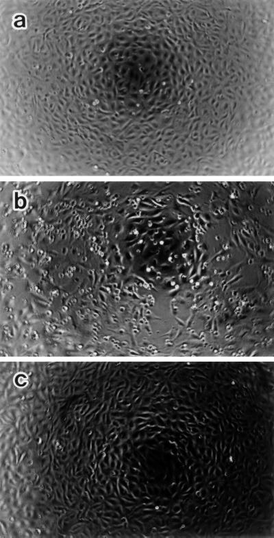
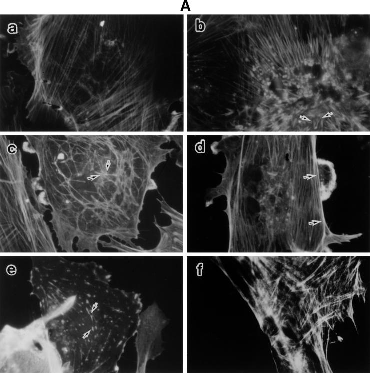
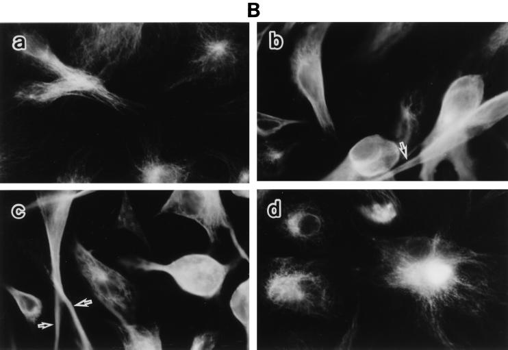
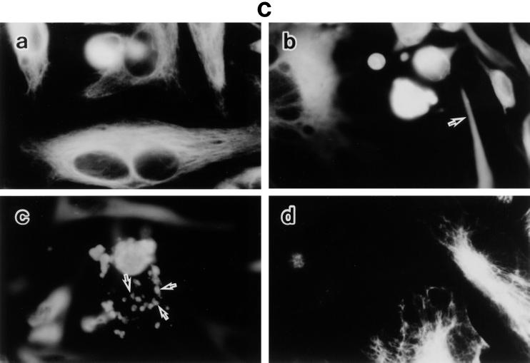
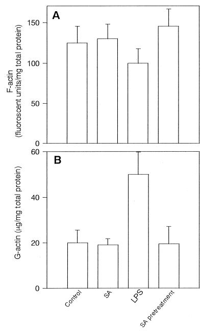
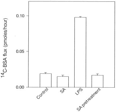
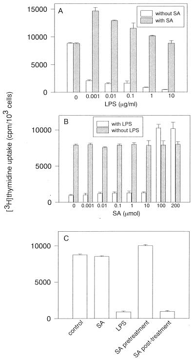
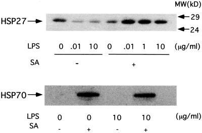
Morphological changes, especially cytoskeletal alterations, in lipopolysaccharide (LPS)-induced vascular endothelial cell injury were studied by using LPS-susceptible bovine aortic endothelial cells (BAEC). BAEC in cultures with LPS showed cell rounding, shrinking, and intercellular gap formation. In those cells, LPS caused the disorganization of actin, tubulin, and vimentin. LPS also induced a reduction in the F-actin pool and an elevation in the G-actin pool. Cytoskeletal disorganization affected transendothelial permeability across the endothelial monolayer. Pretreatment of BAEC with sodium arsenite (SA) prevented alterations in LPS-induced BAEC injury. However, posttreatment with SA had no protective effect on them. SA upregulated the expression of heat shock protein in the presence of LPS. The role of SA in prevention of LPS-induced BAEC injury is discussed.


Figures

FIG. 1
FIG. 1
Phase-contrast micrographs showing BAEC morphology. Untreated (a and b) and SA-pretreated (c) BAEC were cultured with medium alone (a) or with LPS (10 μg/ml) (b and c) for 10 h. Note that LPS induces cell shrinking, rounding, and intercellular gaps in BAEC (b), but not in SA-pretreated BAEC (c). Original magnification, ×25.
FIG. 2
FIG. 2
LPS-induced disorganization of F-actin, tubulin, and vimentin and its prevention by SA pretreatment. Fluorescent micrographs show F-actin (A), tubulin (B), and vimentin (C) organization in response to LPS. Untreated and SA-pretreated BAEC were cultured with various concentrations of LPS for 6 h. (A) For estimation of F-actin organization, BAEC were cultured with medium alone (a) or 0.01 (b), 0.1 (c), 1 (d), or 10 (e) μg of LPS per ml. SA-pretreated BAEC (f) were cultured with 10 μg of LPS per ml. Arrows indicate the extensive spike formation of F-actin (b), its peripheral accumulation (c and d), and the knotted form of actin staining (e). (B) For estimation of tubulin organization, BAEC were cultured with medium alone (a) or 0.01 (b) or 10 (c) μg of LPS per ml. SA-pretreated BAEC (d) were cultured with 10 μg of LPS per ml. Arrows indicate loss of the filamentous network. (C) The same samples were used for the staining of vimentin organization. Arrows in panels b and c show homogenous vimentin staining and cell blebs and round bodies surrounding the cell, respectively. Original magnification, ×500.
FIG. 2
FIG. 2
LPS-induced disorganization of F-actin, tubulin, and vimentin and its prevention by SA pretreatment. Fluorescent micrographs show F-actin (A), tubulin (B), and vimentin (C) organization in response to LPS. Untreated and SA-pretreated BAEC were cultured with various concentrations of LPS for 6 h. (A) For estimation of F-actin organization, BAEC were cultured with medium alone (a) or 0.01 (b), 0.1 (c), 1 (d), or 10 (e) μg of LPS per ml. SA-pretreated BAEC (f) were cultured with 10 μg of LPS per ml. Arrows indicate the extensive spike formation of F-actin (b), its peripheral accumulation (c and d), and the knotted form of actin staining (e). (B) For estimation of tubulin organization, BAEC were cultured with medium alone (a) or 0.01 (b) or 10 (c) μg of LPS per ml. SA-pretreated BAEC (d) were cultured with 10 μg of LPS per ml. Arrows indicate loss of the filamentous network. (C) The same samples were used for the staining of vimentin organization. Arrows in panels b and c show homogenous vimentin staining and cell blebs and round bodies surrounding the cell, respectively. Original magnification, ×500.
FIG. 3
FIG. 3
LPS-induced alteration of the F- and G-actin pools and its prevention by SA pretreatment. F-actin (A) and G-actin (B) pools were determined in untreated and SA-pretreated BAEC 6 h after cultivation with LPS (100 ng/ml). The F-actin pool is expressed as the mean fluorescent units per milligram of total cell protein of triplicates ± standard deviation in three independent experiments. G-actin in the same samples was assayed by DNase I inhibition assay, and each bar represents the mean G-actin in micrograms per milligram of total cell protein of triplicates ± standard deviation in three independent experiments.
FIG. 4
FIG. 4
Effect of pretreatment or posttreatment with SA on transendothelial 14C-BSA flux in cultures of BAEC with LPS. SA-pretreated BAEC were cultured with LPS (100 ng/ml) for 6 h. For posttreatment with LPS, BAEC were cultured with LPS for 6 h and further incubated with SA for 90 min, followed by washing. Transendothelial flux was determined by cultivation of BAEC with 14C-BSA for 1 h. Each bar represents the mean 14C-BSA flux (picomoles per hour) of triplicates ± standard deviation in three independent experiments.
FIG. 5
FIG. 5
LPS-induced reduction of [3H]thymidine incorporation in BAEC and its prevention by SA. (A) Effect of SA on [3H]thymidine incorporation in cultures of BAEC with addition of various concentrations of LPS. SA-pretreated BAEC were cultured with various concentrations of LPS for 24 h and further incubated with [3H]thymidine for 18 h. (B) Effect of pretreatment with various concentrations of SA on [3H]thymidine incorporation in the presence or absence of LPS (1 μg/ml). (C) Effect of pretreatment or posttreatment with SA on [3H]thymidine incorporation in cultures of BAEC with LPS. SA-pretreated BAEC were cultured with LPS (10 μg/ml) for 8 h. Posttreatment with SA was performed after cultivation of BAEC with LPS for 8 h. BAEC were further incubated with [3H]thymidine for 18 h. [3H]thymidine incorporation for DNA synthesis is expressed as the mean counts per minute of triplicates ± standard deviation in five independent experiments.
FIG. 6
FIG. 6
Immunoblotting analysis of HSP27 and HSP70 expression. Untreated and SA-pretreated BAEC were cultured with various concentrations of LPS for 6 h. Extracts from cells treated with 10 μg of LPS per ml or those treated with 0.01, 0.1, 1, and 10 μg of LPS per ml were analyzed by immunoblotting. MW, molecular mass.

Similar articles

Cited by

References

    1. Alexander J S, Hechtman H B, Shepro D. Phalloidin enhances endothelial barrier function and reduces inflammatory permeability in vitro. Microvasc Res. 1988;35:308–315. - PubMed
    1. Benndorf R, Hayess K, Ryazantsev S, Wieske M, Behlke J, Lutsch G. Phosphorylation and supramolecular organization of murine small heat shock protein HSP25 abolish its actin polymerizing-inhibiting activity. J Biol Chem. 1994;269:20780–20784. - PubMed
    1. Brandtzaeg P, Kierulf P, Gaustad P, Skulberg A, Bruun J N, Halversen S, Sorensen E. Plasma endotoxin as a predictor of multiple organ failure and death in systemic meningococcal disease. J Infect Dis. 1989;159:195–204. - PubMed
    1. Brostrom C O, Brostrom M A. Regulation of translational initiation during cellular responses to stress. Prog Nucleic Acid Res Mol Biol. 1998;58:79–125. - PubMed
    1. Burdon R H. Temperature and animal cell protein synthesis. Symp Soc Exp Biol. 1987;41:113–133. - PubMed

Publication types

MeSH terms

LinkOut - more resources