. 2000 Mar 6;148(5):849-56.
doi: 10.1083/jcb.148.5.849.

Nuclear export of MAP kinase (ERK) involves a MAP kinase kinase (MEK)-dependent active transport mechanism

Affiliations

Nuclear export of MAP kinase (ERK) involves a MAP kinase kinase (MEK)-dependent active transport mechanism

M Adachi et al. J Cell Biol. .

Erratum in

  • J Cell Biol 2000 May 1;149(3):754

Abstract

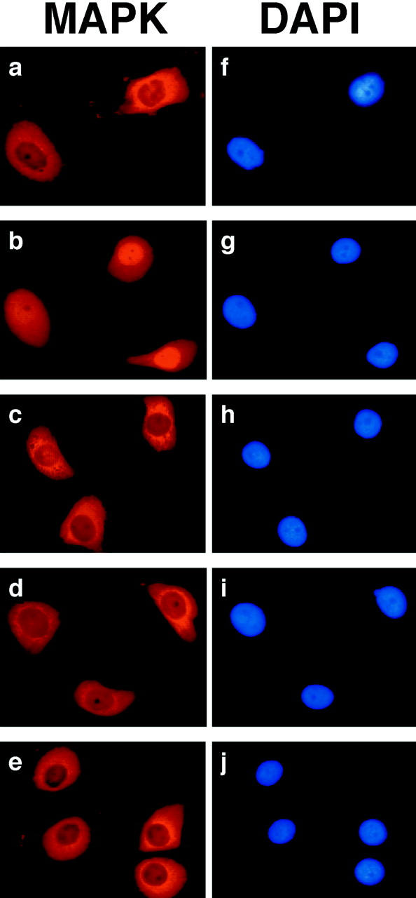
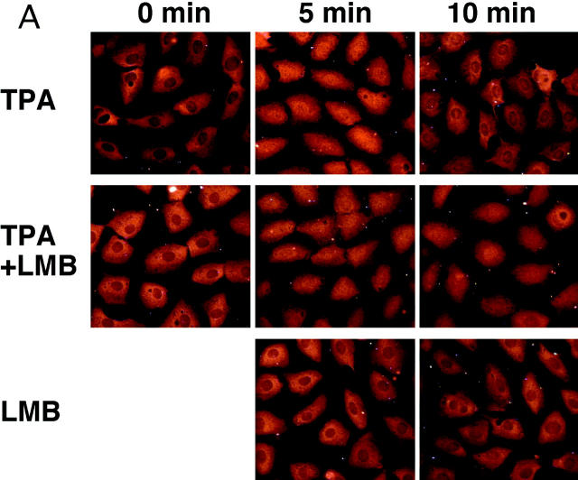
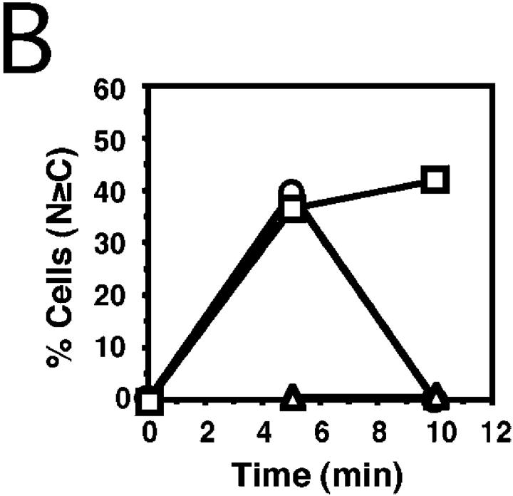
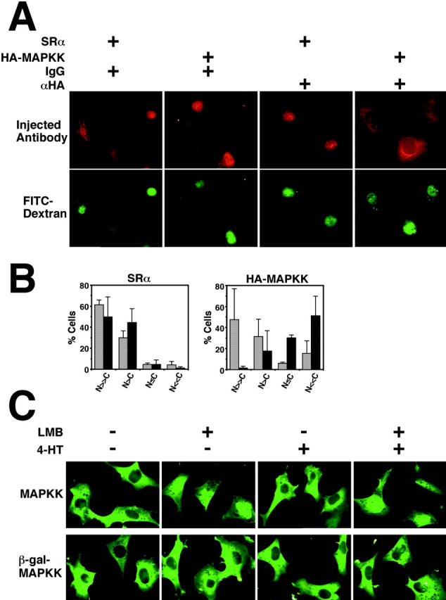
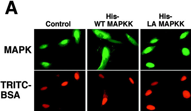
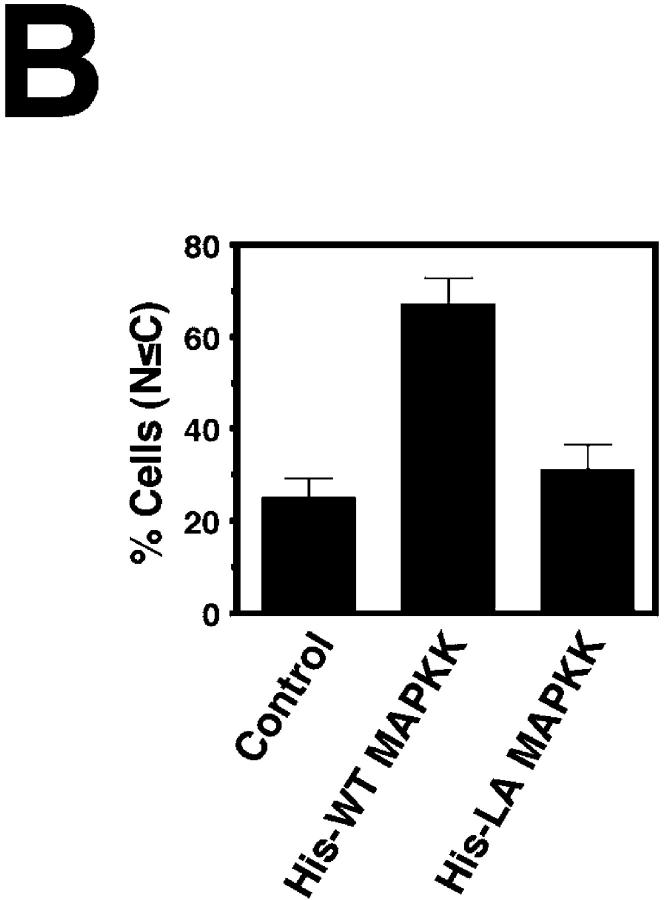
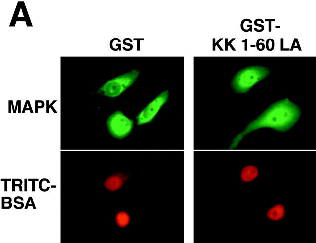
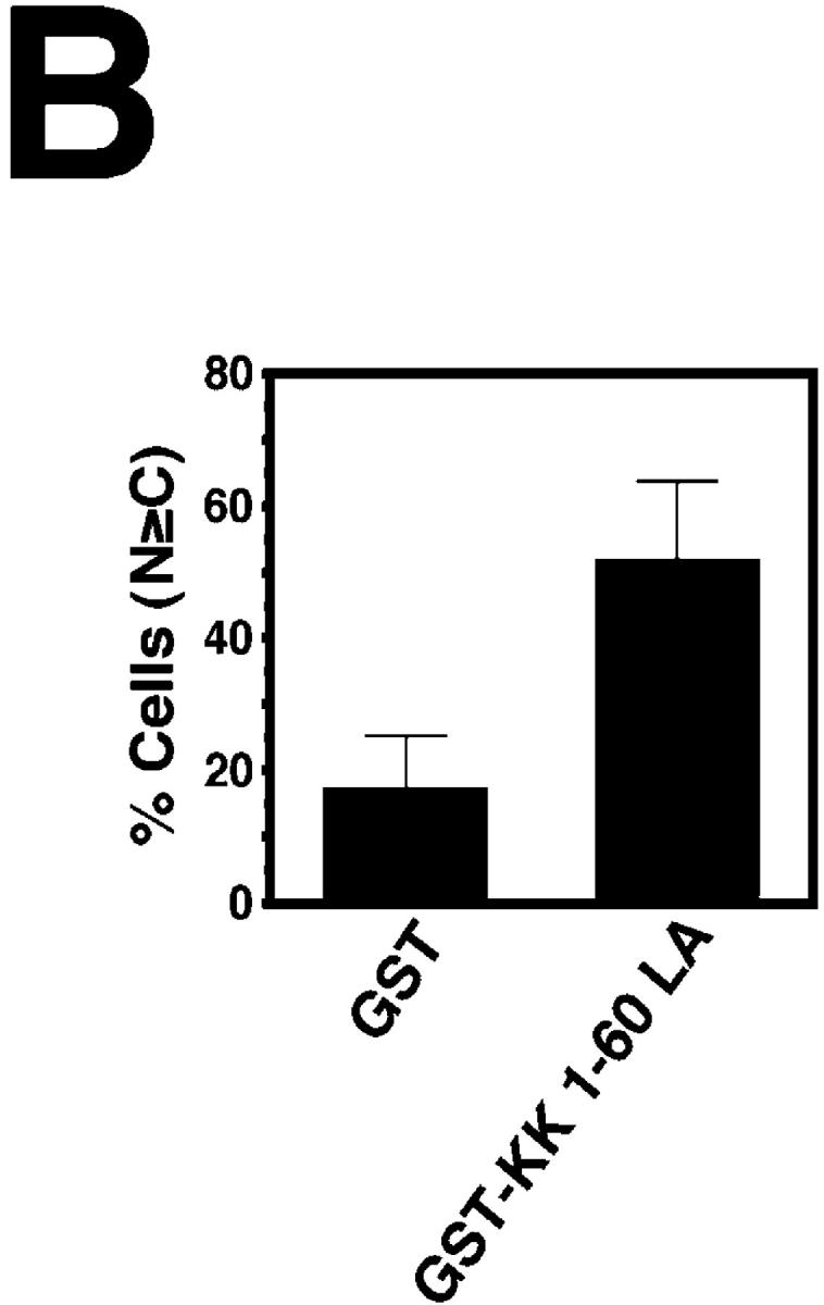
In response to extracellular stimuli, mitogen-activated protein kinase (MAPK, also known as ERK), which localizes to the cytoplasm in quiescent cells, translocates to the nucleus and then relocalizes to the cytoplasm again. The relocalization of nuclear MAPK to the cytoplasm was not inhibited by cycloheximide, confirming that the relocalization is achieved by nuclear export, but not synthesis, of MAPK. The nuclear export of MAPK was inhibited by leptomycin B (LMB), a specific inhibitor of the nuclear export signal (NES)-dependent transport. We have then shown that MAP kinase kinase (MAPKK, also known as MEK), which mostly localizes to the cytoplasm because of its having NES, is able to shuttle between the cytoplasm and the nucleus constantly. MAPK, when injected into the nucleus, was rapidly exported from the nucleus by coinjected wild-type MAPKK, but not by the NES-disrupted MAPKK. In addition, injection of the fragment corresponding to the MAPK-binding site of MAPKK into the nucleus, which would disrupt the binding of MAPK to MAPKK in the nucleus, significantly inhibited the nuclear export of endogenous MAPK. Taken together, these results suggest that the relocalization of nuclear MAPK to the cytoplasm involves a MAPKK-dependent, active transport mechanism.


Figures

Figure 1
Figure 1
Relocalization of nuclear MAPK to the cytoplasm is mediated by nuclear export. Staining images of cells with anti-Xenopus MAPK antibody (a–e) or DAPI (f–j) are shown. A6 cells were serum starved for 36 h (a and f), and then stimulated with 10% FCS for 4 h (b and g). Then cells were treated with CHX (50 μg/ml) for 10 min, followed by deprivation of serum for 10 min in the presence of CHX (d and i), or cells were treated with control buffer for 10 min and then deprived of serum for 10 min in the presence of the buffer (c and h). Incubation of cells with CHX or buffer for 10 min alone did not change subcellular distribution of MAPK (data not shown). In agreement with the result of Lenormand et al. 1998, stimulation of cells with 10% FCS for 4 h in the presence of CHX (50 μg/ml) did not induce nuclear accumulation of MAPK (e and j), indicating that CHX at this concentration is effective. Experiments were performed twice with similar results.
Figure 2
Figure 2
LMB inhibits the nuclear export of MAPK. 3Y1 cells were serum starved for 36 h. Then, the cells were stimulated with TPA alone (500 ng/ml, upper panels in A, circles in B). LMB (0.4 ng/ml) was added 5 min after the addition of TPA (middle panels in A, squares in B), or only LMB without TPA treatment was added (lower panels in A, triangles in B). Cells were fixed, and then stained with anti-ERK1 antibody. The representative images are shown in A. (B) Quantification of the data from a representative experiment. The percentages of cells in which nuclear MAPK staining was stronger than, or equal to, cytoplasmic MAPK staining are shown in each condition. 86–116 cells were examined in each condition. Experiments were performed four times with similar results.
Figure 2
Figure 2
LMB inhibits the nuclear export of MAPK. 3Y1 cells were serum starved for 36 h. Then, the cells were stimulated with TPA alone (500 ng/ml, upper panels in A, circles in B). LMB (0.4 ng/ml) was added 5 min after the addition of TPA (middle panels in A, squares in B), or only LMB without TPA treatment was added (lower panels in A, triangles in B). Cells were fixed, and then stained with anti-ERK1 antibody. The representative images are shown in A. (B) Quantification of the data from a representative experiment. The percentages of cells in which nuclear MAPK staining was stronger than, or equal to, cytoplasmic MAPK staining are shown in each condition. 86–116 cells were examined in each condition. Experiments were performed four times with similar results.
Figure 3
Figure 3
MAPKK shuttles between the cytoplasm and the nucleus. (A and B) 3Y1 cells were serum starved for 48 h. Then, the nuclei were injected with anti-HA antibody (αHA, 20.0 mg/ml) or control IgG (IgG, 20.4 mg/ml) together with the plasmid harboring HA-tagged MAPKK (HA-MAPKK, 150 μg/ml) or the corresponding amount of an empty vector (SRα), all together with FITC-labeled dextran (FITC-Dextran, 800 μg/ml). 5 or 16 h after injection, cells were fixed and stained with Cy3-labeled anti–mouse IgG. The typical images at 16 h after injection are shown in A. (B) Quantification of the data from two independent experiments for nuclear injection of anti-HA antibody (αHA) with SRα or the HA-MAPKK plasmid. The cells were classified into four categories in terms of location of the injected αHA antibody (= Cy3 staining intensity): N >> C, staining intensity in the nucleus (N) is much stronger than that in the cytoplasm (C); N > C, N is stronger than C; N ≤ C, C is equal to or stronger than N; N << C, C is much stronger than N (the αHA is almost completely exported from the nucleus). 56–295 cells were examined in each condition in one experiment. Grey bars, 5 h after injection; black bars, 16 h after injection. Under the conditions used, expression of HA-MAPKK, which was revealed by anti-HA staining in another series of experiments, occurred in almost all the plasmid-injected cells within 4 h. The export of the nuclear injected αHA from the nucleus was clearly detected even 5 h after the nuclear injection of the HA-MAPKK plasmid (see this figure), so the shuttling of HA-MAPKK may occur frequently. (C) ΔB-Raf:ER cells (Pritchard et al. 1995) were transfected with either SRαHA-MAPKK or SRαHA-β-gal-MAPKK together with pCDNA3 MAPK. 16 h later cells were treated with LMB (LMB, 20 ng/ml) and/or 4-hydroxytamoxifen (4-HT, 1 μM), which activates ΔB-Raf, for 5 h. Cells were then fixed and stained with anti-HA antibody. Experiments were performed twice with similar results.
Figure 4
Figure 4
Nuclear export of MAPK protein is induced by nuclear coinjection of wild-type MAPKK, but not by that of NES-disrupted MAPKK. The nuclei of 3Y1 cells were injected with TRITC-BSA and His-tagged MAPK protein (5.0 mg/ml) without (Control) or with His-tagged wild-type MAPKK protein (5.0 mg/ml; His-WT MAPKK) or His-tagged, NES-disrupted MAPKK protein (5.0 mg/ml; His-LA MAPKK). 5 min after injection, cells were fixed and stained with anti-MAPK antibody. The representative images are shown in A. (B) Quantification of the data from three independent experiments. The percentages of cells which showed stronger MAPK fluorescence intensity in the cytoplasm than in the nucleus are shown in each condition. 76–272 cells were examined in each condition in one experiment.
Figure 4
Figure 4
Nuclear export of MAPK protein is induced by nuclear coinjection of wild-type MAPKK, but not by that of NES-disrupted MAPKK. The nuclei of 3Y1 cells were injected with TRITC-BSA and His-tagged MAPK protein (5.0 mg/ml) without (Control) or with His-tagged wild-type MAPKK protein (5.0 mg/ml; His-WT MAPKK) or His-tagged, NES-disrupted MAPKK protein (5.0 mg/ml; His-LA MAPKK). 5 min after injection, cells were fixed and stained with anti-MAPK antibody. The representative images are shown in A. (B) Quantification of the data from three independent experiments. The percentages of cells which showed stronger MAPK fluorescence intensity in the cytoplasm than in the nucleus are shown in each condition. 76–272 cells were examined in each condition in one experiment.
Figure 5
Figure 5
Nuclear export of MAPK is significantly inhibited by inhibiting its binding to MAPKK in the nucleus. A6 cells were serum starved for 36 h, and stimulated with 10% FCS for 4 h. Then, the nuclei were injected with GST (15.0 mg/ml) or the GST fusion protein of the NH2-terminal 1–60 residues of MAPKK with a disrupted NES (GST-KK 1-60 LA, 16.0 mg/ml) together with TRITC-BSA. Then, the culture medium was replaced by the serum-free medium. 10 min after the serum removal, cells were fixed and stained with anti-MAPK antibody. The typical images are shown in A. (B) Quantification of the data from three independent experiments. The percentages of cells which showed stronger MAPK staining in the nucleus than in the cytoplasm are shown in each condition. 133–262 cells were examined in each condition in one experiment.
Figure 5
Figure 5
Nuclear export of MAPK is significantly inhibited by inhibiting its binding to MAPKK in the nucleus. A6 cells were serum starved for 36 h, and stimulated with 10% FCS for 4 h. Then, the nuclei were injected with GST (15.0 mg/ml) or the GST fusion protein of the NH2-terminal 1–60 residues of MAPKK with a disrupted NES (GST-KK 1-60 LA, 16.0 mg/ml) together with TRITC-BSA. Then, the culture medium was replaced by the serum-free medium. 10 min after the serum removal, cells were fixed and stained with anti-MAPK antibody. The typical images are shown in A. (B) Quantification of the data from three independent experiments. The percentages of cells which showed stronger MAPK staining in the nucleus than in the cytoplasm are shown in each condition. 133–262 cells were examined in each condition in one experiment.

References

    1. Adachi M., Fukuda M., Nishida E. Two co-existing mechanisms for nuclear import of MAP kinasepassive diffusion of a monomer and active transport of a dimer. EMBO (Eur. Mol. Biol. Organ.) J. 1999;18:5347–5358. - PMC - PubMed
    1. Bardwell L., Cook J.G., Chang E.C., Cairns B.R., Thorner J. Signaling in the yeast pheromone response pathwayspecific and high-affinity interaction of the mitogen-activated protein (MAP) kinases Kss1 and Fus3 with the upstream MAP kinase kinase Ste7. Mol. Cell. Biol. 1996;16:3637–3650. - PMC - PubMed
    1. Blenis J. Signal transduction via the MAP kinaseproceed at your own RSK. Proc. Natl Acad. Sci. USA. 1993;90:5889–5892. - PMC - PubMed
    1. Chen R.-H., Sarnecki C., Blenis J. Nuclear localization and regulation of erk- and rsk-encoded protein kinases. Mol. Cell. Biol. 1992;12:915–927. - PMC - PubMed
    1. Davis R.J. The mitogen-activated protein kinase signal transduction pathway. J. Biol. Chem. 1993;268:14553–14556. - PubMed

Publication types

MeSH terms