. 2000 Mar 6;148(5):931-44.
doi: 10.1083/jcb.148.5.931.

PEX19 binds multiple peroxisomal membrane proteins, is predominantly cytoplasmic, and is required for peroxisome membrane synthesis

Affiliations

PEX19 binds multiple peroxisomal membrane proteins, is predominantly cytoplasmic, and is required for peroxisome membrane synthesis

K A Sacksteder et al. J Cell Biol. .

Abstract

Peroxisomes are components of virtually all eukaryotic cells. While much is known about peroxisomal matrix protein import, our understanding of how peroxisomal membrane proteins (PMPs) are targeted and inserted into the peroxisome membrane is extremely limited. Here, we show that PEX19 binds a broad spectrum of PMPs, displays saturable PMP binding, and interacts with regions of PMPs required for their targeting to peroxisomes. Furthermore, mislocalization of PEX19 to the nucleus leads to nuclear accumulation of newly synthesized PMPs. At steady state, PEX19 is bimodally distributed between the cytoplasm and peroxisome, with most of the protein in the cytoplasm. We propose that PEX19 may bind newly synthesized PMPs and facilitate their insertion into the peroxisome membrane. This hypothesis is supported by the observation that the loss of PEX19 results in degradation of PMPs and/or mislocalization of PMPs to the mitochondrion.


Figures
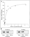
Figure 2
Figure 2
PEX19-PMP binding is saturable. (A) Purified 6xHis-PEX19 was separated by SDS-PAGE, transferred to PVDF membranes, and incubated with purified recombinant 6xHis-PEX14 spiked with 35S-labeled 6xHis-PEX14 at various concentrations, washed, and analyzed using a PhosphorImager. The amount of PEX14 bound at each concentration was determined and plotted versus 6xHis-PEX14 concentration using KaleidaGraph software. (B) Membranes containing 1 μg of immobilized 6xHis-PEX19 were incubated with 35S-PEX14 (left) or 35S-PEX3 (right) in buffer A (left lanes), buffer A containing 10 μM 6xHis-PTE1 (middle lanes), and buffer A containing 10 μM 6xHis-PEX14 (right lanes).
Figure 3
Figure 3
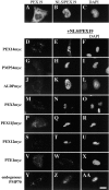
Targeting PEX19 to the nucleus leads to nuclear accumulation of PMPs. (A–C) Human fibroblasts expressing PEX19 (A) or NLS/PEX19 (B and C) were analyzed by indirect immunofluorescence using affinity-purified anti–PEX19 antibodies (A and B) and stained with DAPI (C). Human fibroblasts expressing the myc-tagged PMPs PEX14 (D–F), PMP34 (G–I), ALDP (J–L), PEX3 (M–O), PEX11β (P–R), or PEX12 (S–U), or the peroxisomal matrix protein PTE1 (V–X) with pcDNA3 alone (left column) or with pcDNA3-NLS/PEX19 (middle and right columns) were analyzed by indirect immunofluorescence using monoclonal anti–myc antibodies with appropriate secondary antibodies (left and middle columns) and DAPI (right column). Fibroblasts transfected with pcDNA3 (Y) or pcDNA3-NLS/PEX19 (Z and AA) were analyzed by indirect immunofluorescence using anti–PMP70 antibodies (Y and Z) with appropriate secondary antibodies. Cells expressing NLS/PEX19 were also stained with DAPI (AA). Bar, 15 μm.
Figure 4
Figure 4
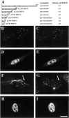
PEX19 binds the peroxisomal targeting element of PMP70. (A) Line diagram of PMP70 mutants. Full-length PMP70 is shown at the top, and predicted transmembrane domains are shown in black boxes. Subcellular distribution and interaction with PEX19 are indicated to the right. Normal human fibroblasts expressing the PMP70 mutant constructs ΔC535PMP70myc (B–E) or ΔC598PMP70myc (F–I) were cotransfected with either vector alone (B, C, F, and G) or pcDNA3-NLS/PEX19 (D, E, H, and I). The distribution of the PMP70 truncation mutants was determined by immunofluorescence using anti–myc antibodies (left). Cells cotransfected with vector alone were stained with antibodies to the COOH terminus of PMP70 (C and G), whereas cells cotransfected with pcDNA3-NLS/PEX19 were stained with DAPI (E and I). Bar, 15 μM.
Figure 5
Figure 5
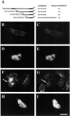
PEX19 binds the peroxisomal targeting element of PEX11β. (A) Line diagram of PEX11 (truncations, their subcellular distribution, and their ability to interact with PEX19 in the nuclear localization assay). Solid boxes show the position of the two transmembrane domains. (B–I) Subcellular distribution of two truncated forms of PEX11β in normal cells and in cells expressing NLS/PEX19. Normal human fibroblasts were transfected with pcDNA3-ΔN180/PEX11 (B-E) or pcDNA3-ΔN210/PEX11β (F–I) and cotransfected with a vector control (B, C, F, and G) or pcDNA3-NLS/PEX19 (D, E, H, and I). Cells were processed for indirect immunofluorescence using antibodies specific for the myc epitope tag (B, D, F, and H) and colabeled with antibodies specific for endogenously expressed PMP70 (C and G) or DAPI (E and I). Bar, 15 μm.
Figure 6
Figure 6
Analysis of PEX14 targeting and interaction with PEX19. (A) Line diagram of PEX14 truncation mutations. Solid boxes show the position of the transmembrane domain. (B–G) Normal human fibroblasts expressing PEX14myc (B and C), ΔC231PEX14myc (D and E), or ΔC270PEX14myc (F and G) were processed for indirect immunofluorescence using antibodies specific for the myc epitope tag (left) and PMP70 (right) using appropriate secondary antibodies. (H) Competition of PEX14-PEX19 binding by WT and mutant PEX14 proteins. Purified 6xHis-PEX19 was separated by SDS-PAGE, transferred to PVDF membranes, and probed with equal amounts of 35S-labeled PEX14 in buffer A or buffer A containing various concentrations of 6xHis-PEX14, 6xHis-ΔC231PEX14, and 6xHis-ΔC270PEX14. Membranes were washed and the amount of 35S-labeled PEX14 bound to PEX19 was determined using a PhosphorImager. These values were normalized to the control reaction and plotted on a bar graph. Bar, 15 μM.
Figure 7
Figure 7
Subcellular distribution of mammalian PEX19. (A) A postnuclear supernatant of rat liver was fractionated by centrifugation on a 15–45% Nycodenz gradient and the position of the peroxisomal marker (catalase, solid line), the mitochondrial marker (succinate dehydrogenase, dotted line), and a cytoplasmic marker (lactate dehydrogenase, dashed line) are shown. The bottom of the gradient (most dense) is to the left, and the abundance of PEX19 in each fraction was determined by immunoblot (lower panel, arrow). (B) Highly purified rat liver peroxisomes were separated by SDS-PAGE and PEX19 was detected by immunoblot (arrow). (C) Rat liver was homogenized and subjected to differential centrifugation analysis. Equal proportions of homogenate, 25,000 g pellet, 25,000 g supernatant, 100,000 g pellet, and 100,000 g supernatant fractions were separated by SDS-PAGE and assayed by immunoblot for PEX19. (D) Human fibroblasts (GM5756-T) were harvested and incubated with varying amounts of digitonin, as well as digitonin and Triton X-100. Insoluble material was pelleted by centrifugation, and the supernatants were assayed for the release of cytoplasmic (LDH, square) and peroxisomal (catalase, circle) markers and for PEX19 by immunoblot (diamond). (E–G) Fibroblasts were transfected with pcDNA3 (E), pcDNA3-PEX19 (F), or pcDNA3-PEX19/C296A (G) and processed by immunofluorescence using antibodies to catalase and the appropriate secondary antibodies.
Figure 8
Figure 8
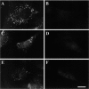
A subset of PMPs is not detectable in PEX19-deficient cells. Wild-type (left column) and PBD399 (right column) human fibroblasts were processed for indirect immunofluorescence using antibodies specific for PMP70 (A and B), PEX13 (C and D), and PEX11β (E and F) with the appropriate secondary antibodies. Bar, 15 μm.
Figure 9
Figure 9
A subset of PMPs localizes to mitochondria in PEX19-deficient cells. The distribution of endogenous PEX14 was determined in wild-type (A) and PBD399 (B) fibroblasts using affinity-purified PEX-14 antibodies. The distribution of PEX12myc (D and E), ALDPmyc (G and H), and PEX3myc (J and K) was determined in wild-type (D, G, and J) and PBD399 (E, H, and K) fibroblasts using antibodies specific to the myc epitope. PBD399 fibroblasts were also stained with MitoTracker (C, F, I, and L). The peroxisomal nature of the punctate structures in wild-type fibroblasts was confirmed by double label with anti–PMP70 antibodies (data not shown). Bar, 15 μm.

Similar articles

Cited by

References

    1. Adams G.A., Rose J.K. Structural requirements of a membrane-spanning domain for protein anchoring and cell surface transport. Cell. 1985;41:1007–1015. - PubMed
    1. Bjorkman J., Stetten G., Moore C.S., Gould S.J., Crane D.I. Genomic structure of PEX13, a candidate peroxisome biogenesis disorder gene. Genomics. 1998;54:521–528. - PubMed
    1. Boursnell M.E., Brown T.D., Binns M.M. Sequence of the membrane protein gene from avian coronavirus IBV. Virus Res. 1984;1:303–313. - PMC - PubMed
    1. Braverman N., Dodt G., Gould S.J., Valle D. Disorders of peroxisome biogenesis. Hum. Mol. Genet. 1995;4:1791–1798. - PubMed
    1. Chang C.-C., Gould S.J. Phenotype-genotype relationships in complementation group 3 of the peroxisome-biogenesis disorders. Am. J. Hum. Genet. 1998;63:1294–1306. - PMC - PubMed

Publication types

MeSH terms