

Massive idiosyncratic exon skipping corrects the nonsense mutation in dystrophic mouse muscle and produces functional revertant fibers by clonal expansion
- PMID: 10704448
- PMCID: PMC2174546
- DOI: 10.1083/jcb.148.5.985
Massive idiosyncratic exon skipping corrects the nonsense mutation in dystrophic mouse muscle and produces functional revertant fibers by clonal expansion
Abstract
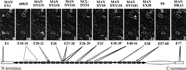
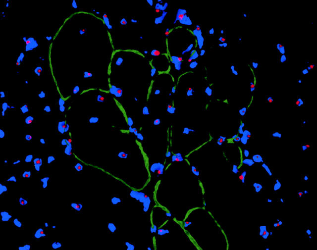
Conventionally, nonsense mutations within a gene preclude synthesis of a full-length functional protein. Obviation of such a blockage is seen in the mdx mouse, where despite a nonsense mutation in exon 23 of the dystrophin gene, occasional so-called revertant muscle fibers are seen to contain near-normal levels of its protein product. Here, we show that reversion of dystrophin expression in mdx mice muscle involves unprecedented massive loss of up to 30 exons. We detected several alternatively processed transcripts that could account for some of the revertant dystrophins and could not detect genomic deletion from the region commonly skipped in revertant dystrophin. This, together with exon skipping in two noncontiguous regions, favors aberrant splicing as the mechanism for the restoration of dystrophin, but is hard to reconcile with the clonal idiosyncrasy of revertant dystrophins. Revertant dystrophins retain functional domains and mediate plasmalemmal assembly of the dystrophin-associated glycoprotein complex. Physiological function of revertant fibers is demonstrated by the clonal growth of revertant clusters with age, suggesting that revertant dystrophin could be used as a guide to the construction of dystrophin expression vectors for individual gene therapy. The dystrophin gene in the mdx mouse provides a favored system for study of exon skipping associated with nonsense mutations.
Figures
Similar articles
-
Mutation types and aging differently affect revertant fiber expansion in dystrophic mdx and mdx52 mice.PLoS One. 2013 Jul 24;8(7):e69194. doi: 10.1371/journal.pone.0069194. Print 2013. PLoS One. 2013. PMID: 23894429 Free PMC article.
-
Revertant fibres: a possible genetic therapy for Duchenne muscular dystrophy?Neuromuscul Disord. 1997 Jul;7(5):329-35. doi: 10.1016/s0960-8966(97)00058-8. Neuromuscul Disord. 1997. PMID: 9267847
-
Morpholino-induced exon skipping stimulates cell-mediated and humoral responses to dystrophin in mdx mice.J Pathol. 2019 Jul;248(3):339-351. doi: 10.1002/path.5263. Epub 2019 Apr 16. J Pathol. 2019. PMID: 30883742 Free PMC article.
-
Skipping multiple exons of dystrophin transcripts using cocktail antisense oligonucleotides.Nucleic Acid Ther. 2014 Feb;24(1):57-68. doi: 10.1089/nat.2013.0451. Epub 2013 Dec 31. Nucleic Acid Ther. 2014. PMID: 24380394 Review.
-
Screening for antisense modulation of dystrophin pre-mRNA splicing.Neuromuscul Disord. 2002 Oct;12 Suppl 1:S67-70. doi: 10.1016/s0960-8966(02)00085-8. Neuromuscul Disord. 2002. PMID: 12206799 Review.
Cited by
-
Fetal microchimeric cells in a fetus-treats-its-mother paradigm do not contribute to dystrophin production in serially parous mdx females.Stem Cells Dev. 2012 Oct 10;21(15):2809-16. doi: 10.1089/scd.2012.0047. Epub 2012 Aug 6. Stem Cells Dev. 2012. PMID: 22731493 Free PMC article.
-
Therapeutic Potential of Immunoproteasome Inhibition in Duchenne Muscular Dystrophy.Mol Ther. 2016 Nov;24(11):1898-1912. doi: 10.1038/mt.2016.162. Epub 2016 Aug 10. Mol Ther. 2016. PMID: 27506451 Free PMC article.
-
In vivo gene editing in dystrophic mouse muscle and muscle stem cells.Science. 2016 Jan 22;351(6271):407-411. doi: 10.1126/science.aad5177. Epub 2015 Dec 31. Science. 2016. PMID: 26721686 Free PMC article.
-
Proceedings of the 2017 National Toxicology Program Satellite Symposium.Toxicol Pathol. 2017 Oct;45(7):799-833. doi: 10.1177/0192623317733924. Epub 2017 Nov 7. Toxicol Pathol. 2017. PMID: 29113559 Free PMC article.
-
Rapid direct sequence analysis of the dystrophin gene.Am J Hum Genet. 2003 Apr;72(4):931-9. doi: 10.1086/374176. Epub 2003 Mar 11. Am J Hum Genet. 2003. PMID: 12632325 Free PMC article.
References
-
- Allen D.L., Roy R.R., Edgerton V.R. Myonuclear domain in muscle adaptation and disease. Muscle Nerve. 1999;22:1350–1360. - PubMed
-
- Amalfitano A., Rafael J.A., Chamberlain J.S. Structure and mutation of the dystrophin gene. In: Brown S.C., Lucy-Jack A., editors. Dystrophin Gene, Protein and Cell Biology. Cambridge University Press; Cambridge, UK: 1997. pp. 1–26.
-
- Baylies M.K., Bate M., Ruiz Gomez M. Myogenesisa view from Drosophila . Cell. 1998;93:921–927. - PubMed
-
- Bockhold K.J., Rosenblatt J.D., Partridge T.A. Aging normal and dystrophic mouse muscleanalysis of myogenicity in cultures of living single fibers. Muscle Nerve. 1998;21:173–183. - PubMed
Publication types
MeSH terms
Substances
LinkOut - more resources
Full Text Sources
Other Literature Sources
Medical
