. 2000 Mar 6;191(5):859-70.
doi: 10.1084/jem.191.5.859.

Deficiency of the stress kinase p38alpha results in embryonic lethality: characterization of the kinase dependence of stress responses of enzyme-deficient embryonic stem cells

Affiliations

Deficiency of the stress kinase p38alpha results in embryonic lethality: characterization of the kinase dependence of stress responses of enzyme-deficient embryonic stem cells

M Allen et al. J Exp Med. .

Abstract

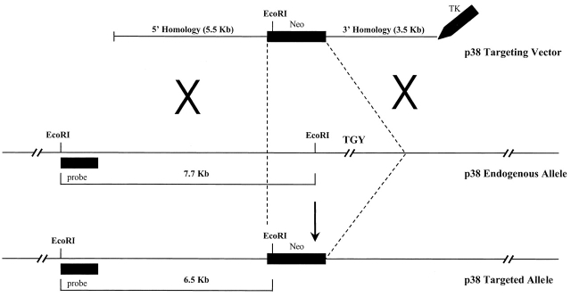
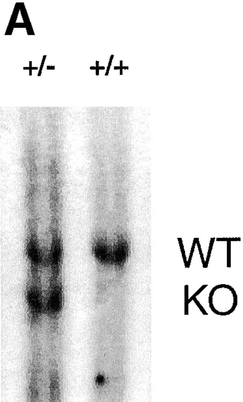
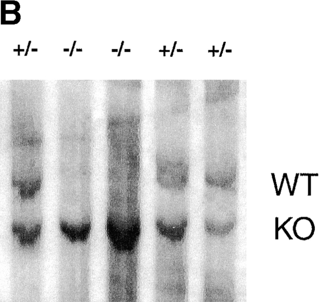
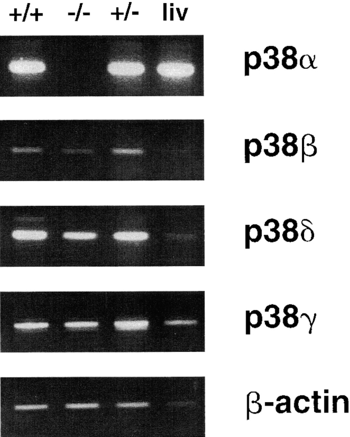
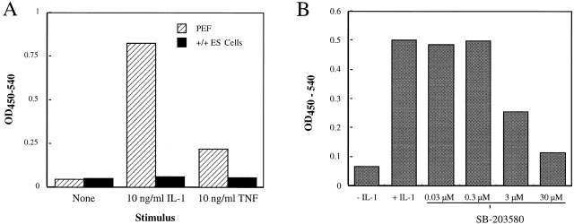
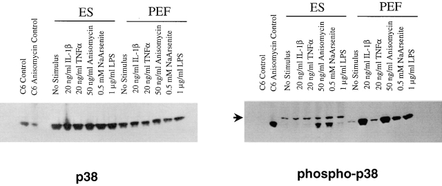
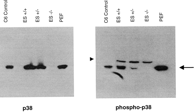
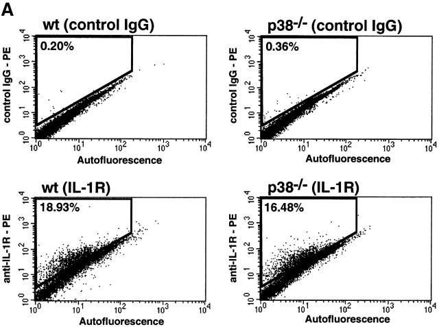
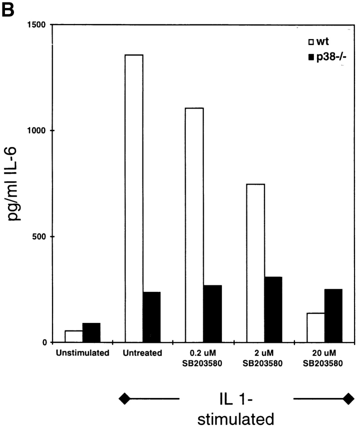
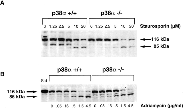
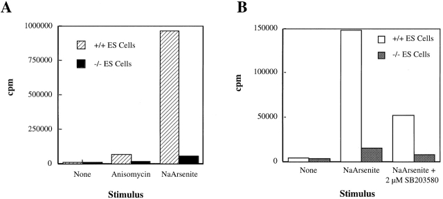
The mitogen-activated protein (MAP) kinase p38 is a key component of stress response pathways and the target of cytokine-suppressing antiinflammatory drugs (CSAIDs). A genetic approach was employed to inactivate the gene encoding one p38 isoform, p38alpha. Mice null for the p38alpha allele die during embryonic development. p38alpha(1/)- embryonic stem (ES) cells grown in the presence of high neomycin concentrations demonstrated conversion of the wild-type allele to a targeted allele. p38alpha(-/)- ES cells lacked p38alpha protein and failed to activate MAP kinase-activated protein (MAPKAP) kinase 2 in response to chemical stress inducers. In contrast, p38alpha(1/+) ES cells and primary embryonic fibroblasts responded to stress stimuli and phosphorylated p38alpha, and activated MAPKAP kinase 2. After in vitro differentiation, both wild-type and p38alpha(-/)- ES cells yielded cells that expressed the interleukin 1 receptor (IL-1R). p38alpha(1/+) but not p38alpha(-/)- IL-1R-positive cells responded to IL-1 activation to produce IL-6. Comparison of chemical-induced apoptosis processes revealed no significant difference between the p38alpha(1/+) and p38alpha(-/)- ES cells. Therefore, these studies demonstrate that p38alpha is a major upstream activator of MAPKAP kinase 2 and a key component of the IL-1 signaling pathway. However, p38alpha does not serve an indispensable role in apoptosis.


Figures

Figure 1
Figure 1
Scheme employed to achieve homologous recombination of the murine p38α locus. Approximately 8–10 kb of endogenous genomic sequence, including regions necessary for p38α enzymatic activity, was replaced by the neomycin gene; this creates a deletion from phenylalanine 129 to aspartic acid 283 within the native protein. Hybridization of ES cell DNA with an external probe identified a predicted RFLP of 6.5 kb, created as a result of the introduction of a novel EcoRI restriction site in the targeting vector.
Figure 2
Figure 2
Southern analysis of ES cell clones. EcoRI restriction enzyme–digested ES cell genomic DNA was electrophoresed, transferred to nitrocellulose, and hybridized to the 5′ external probe of the p38α locus. (A) A blot showing wild-type (+/+) and the parental heterozygous (+/−) ES cell clones. (B) A blot showing examples of G418-surviving ES cell clones; this blot is representative of 64 surviving clones. The positions of the wild-type (WT) and knockout (KO) alleles are indicated.
Figure 2
Figure 2
Southern analysis of ES cell clones. EcoRI restriction enzyme–digested ES cell genomic DNA was electrophoresed, transferred to nitrocellulose, and hybridized to the 5′ external probe of the p38α locus. (A) A blot showing wild-type (+/+) and the parental heterozygous (+/−) ES cell clones. (B) A blot showing examples of G418-surviving ES cell clones; this blot is representative of 64 surviving clones. The positions of the wild-type (WT) and knockout (KO) alleles are indicated.
Figure 3
Figure 3
Expression of the four murine p38 isoforms and β-actin in wild-type (+/+) ES cells, wild-type liver (liv), p38α-deficient (−/−) ES cells, and p38α heterozygous (+/−) ES cells. The observed RT-PCR product sizes agreed with the predicted values and are as follows: p38α, 368 bp; p38β, 430 bp; p38δ, 354 bp; p38γ, 632 bp; and β-actin, 540 bp.
Figure 4
Figure 4
PEFs, but not ES cells, produce IL-6 in response to inflammatory cytokine stimuli. (A) Cultures of PEFs or wild-type ES cells were incubated overnight in medium containing no effector (None) or 10 ng/ml of IL-1β or TNF-α, after which IL-6 released to the medium was measured by ELISA. The ELISA signal is indicated as a function of treatment (average of duplicate determinations). This experiment was repeated twice with comparable results. (B) PEFs were stimulated overnight with 10 ng/ml of IL-1 in the absence or presence of the indicated concentration of SB-203,580. IL-6 released to the medium was determined by ELISA (average of duplicate determinations). These data are representative of three separate experiments.
Figure 5
Figure 5
Identification of stimuli that promote p38α phosphorylation in PEFs and wild-type ES cells. Cells were stimulated for 15 min in the presence of the indicated effector, and then were solubilized by detergent extraction. Equal quantities of these extracts were fractionated on two separate gels, and the separated polypeptides were transferred to nitrocellulose for Western blot analysis. One blot was treated with an antibody that detects total p38α (p38), and the other with an antibody that detects phosphorylated p38α (phospho-p38). Extracts derived from control and anisomycin-treated C6 cells are included as standards. Identity of the slightly slower migrating antigenic species detected in the ES cell extracts with the phosphospecific antibody (arrow) is unknown.
Figure 6
Figure 6
Demonstration that p38α−/− ES cells lack immunodetectable p38α. Wild-type (ES+/+) and p38α-deficient ES cells were stimulated with sodium arsenite for 15 min and then solubilized by detergent extraction. Duplicate samples of each extract were loaded onto separate gels and processed for Western blotting. Antigen detected after staining these blots with an antibody against total (p38) or one selective for phosphospecific (phospho-p38) forms are indicated. Extracts of IL-1–stimulated PEFs and C6 cells are included as standards. Arrow indicates the migration of p38α, and the arrowhead marks the antigen detected by the phospho-specific antiserum in the ES cell samples that is not p38 dependent.
Figure 8
Figure 8
IL-1–induced IL-6 production by in vitro–differentiated ES cells. Wild-type and p38α−/− ES cells were subjected to in vitro differentiation. (A) Cells recovered from each culture were stained with a PE-labeled control IgG or PE-labeled anti–IL-1R IgG, and the cell mixtures were analyzed by FACS®. Cells were gated by autofluorescence, an indicator of size (x-axis), and by PE fluorescence intensity (y-axis). The percentage of cells demonstrating a PE fluorescence intensity above the background level is indicated in each panel. (B) IL-1R–positive cells recovered by sorting were plated into culture wells and stimulated with IL-1β (10 ng/ml) in the absence or presence of SB-203,580. After an overnight stimulation, media were harvested and assayed for IL-6 by ELISA. The amount of IL-6 produced is indicated as a function of treatment. These results are representative of four separate experiments. wt, wild-type.
Figure 8
Figure 8
IL-1–induced IL-6 production by in vitro–differentiated ES cells. Wild-type and p38α−/− ES cells were subjected to in vitro differentiation. (A) Cells recovered from each culture were stained with a PE-labeled control IgG or PE-labeled anti–IL-1R IgG, and the cell mixtures were analyzed by FACS®. Cells were gated by autofluorescence, an indicator of size (x-axis), and by PE fluorescence intensity (y-axis). The percentage of cells demonstrating a PE fluorescence intensity above the background level is indicated in each panel. (B) IL-1R–positive cells recovered by sorting were plated into culture wells and stimulated with IL-1β (10 ng/ml) in the absence or presence of SB-203,580. After an overnight stimulation, media were harvested and assayed for IL-6 by ELISA. The amount of IL-6 produced is indicated as a function of treatment. These results are representative of four separate experiments. wt, wild-type.
Figure 9
Figure 9
Deletion of p38α does not inhibit ES cell apoptosis in response to chemical stimuli. p38α1/+ and p38α−/− ES cells were treated with the indicated concentration of staurosporin (A) or adriamycin (B) for 16 h, and then were disaggregated with SDS sample buffer. Samples of the resulting lysates were subjected to Western blot analysis with an anti-PARP antibody. Regions of the blots corresponding to full-length 116-kD PARP and its 85-kD cleavage fragment are shown as a function of the effector concentration.
Figure 7
Figure 7
MAPKAP kinase 2 activation in wild-type and p38α−/− ES cells. Cultures of p38α1/+ and p38α−/− ES cells were incubated with the indicated effector for 15 min, after which the cells were solubilized by detergent extraction, and MAPKAP kinase 2 was recovered by immunoprecipitation. The resulting immunoprecipitates were then assayed for kinase activity using a peptide substrate and [γ-32P]ATP. Radioactivity (in cpm) incorporated into the peptide substrate is indicated as a function of treatment; a background (no enzyme control) was subtracted to correct for non–peptide-associated radioactivity sticking to the filter. This experiment was repeated twice with comparable results. Each value is the average of duplicate determinations.

References

    1. Karin M. Mitogen-activated protein kinase cascades as regulators of stress responses. Ann. NY Acad. Sci. 1998;851:139–146. - PubMed
    1. Kyriakis J.M., Auruch J. Sounding the alarmprotein kinase cascades activated by stress and inflammation. J. Biol. Chem. 1996;271:24313–24316. - PubMed
    1. Ludwig S., Hoffmeyer A., Goebeler M., Kilian K., Hafner H., Neufeld B., Han J., Rap U.R. The stress inducer arsenite activates mitogen-activated protein kinases extracellular signal-regulated kinases 1 and 2 via a MAPK kinase 6/p38-dependent pathway. J. Biol. Chem. 1998;273:1917–1922. - PubMed
    1. Rouse J., Cohen P., Trigon S., Morange M., Alonso-Liamazares A., Zamanillo D., Hunt T., Nebreda A.R. A novel kinase cascade triggered by stress and heat shock that stimulates MAPKAP kinase-2 and phosphorylation of the small heat shock proteins. Cell. 1994;78:1027–1037. - PubMed
    1. Raingeaud J., Gupta S., Rogers J. S., Dickens M., Han J., Ulevitch R.J., Davis R.J. Pro-inflammatory cytokines and environmental stress cause p38 mitogen-activated protein kinase activation by dual phosphorylation on tyrosine and threonine. J. Biol. Chem. 1995;270:7420–7426. - PubMed

MeSH terms