. 2000 Mar 6;191(5):871-82.
doi: 10.1084/jem.191.5.871.

Cryptococcus neoformans STE12alpha regulates virulence but is not essential for mating

Affiliations

Cryptococcus neoformans STE12alpha regulates virulence but is not essential for mating

Y C Chang et al. J Exp Med. .

Abstract

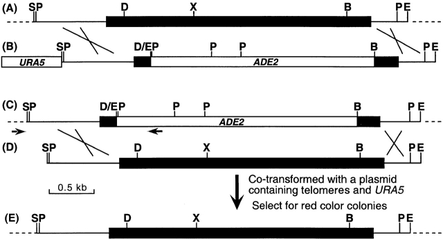
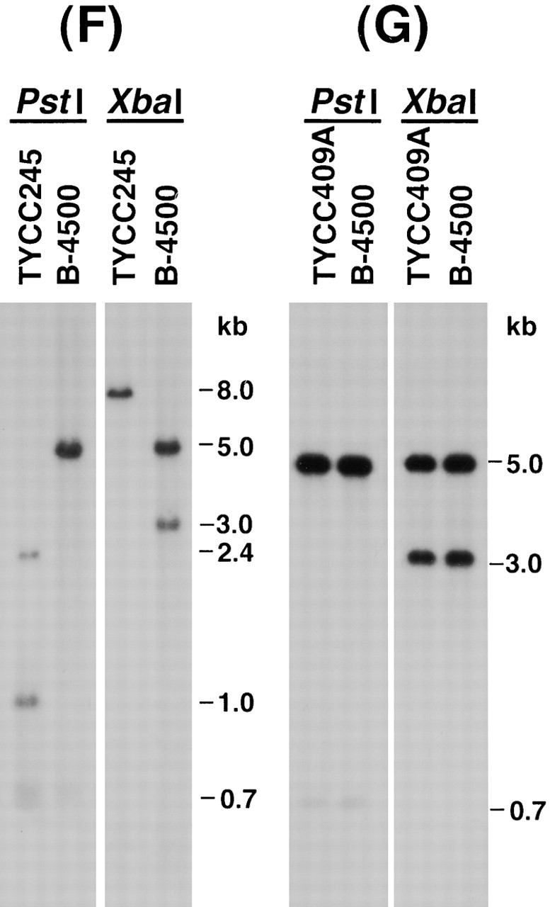
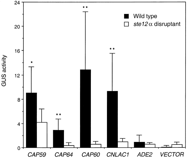
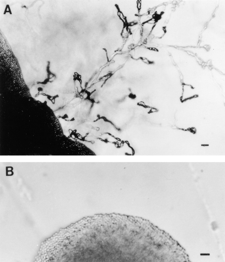
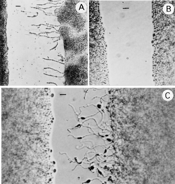
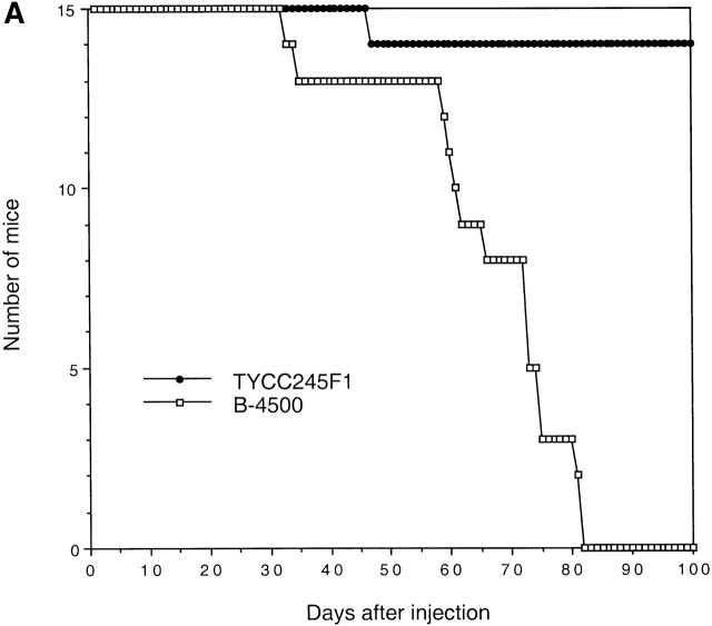
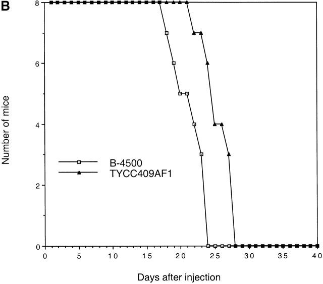
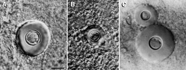
The Cryptococcus neoformans STE12alpha gene, a homologue of Saccharomyces cerevisiae STE12, exists only in mating type (MAT)alpha cells. In S. cerevisiae, STE12 was required for mating and filament formation. In C. neoformans, haploid fruiting on filament agar required STE12alpha. The ability to form hyphae, however, was not affected by deletion of STE12alpha when convergently growing MATa strains were present. Furthermore, ste12alpha disruptants were fertile when mated with MATa strains, albeit with reduced mating frequency. Most importantly, the virulence of a ste12alpha disruptant of serotype D strain was significantly reduced in a mouse model. When the ste12alpha locus was reconstituted with the wild-type allele by cotransformation, virulence was restored. Histopathological analysis demonstrated a reduction in capsular size of yeast cells, less severe cystic lesions, and stronger immune responses in meninges of mice infected with ste12alpha cells than those of mice infected with STE12alpha cells. Using reporter gene constructs, we found that STE12alpha controls the expression of several phenotypes known to be involved in virulence, such as capsule and melanin production. These results demonstrate a clear molecular link between mating type and virulence in C. neoformans.


Figures

Figure 1
Figure 1
(A) Map of the STE12α gene. (B) Map of STE12α deletion construct. For simplicity, the URA5 gene is not drawn to scale. (C) Map of ste12α in TYCC245. (D) Map of DNA fragment used in cotransformation. (E) Map of reconstituted STE12α. Arrowheads, primer locations. Black boxes, coding region of STE12α. Crosses, crossing over. Dashed lines, chromosomal region flanking STE12α. B = BamHI; D = DraIII; E = EcoRI; P = PstI; S = SphI; X = XbaI. (F) Southern blot of ste12α deletant. (G) Southern blot of STE12α reconstituted strain. B-4500, wild type; TYCC245, ste12α deletant; TYCC409A, STE12α reconstituted strain. DNA was isolated, digested with restriction enzymes, fractionated on 0.8% agarose gel, and analyzed by Southern blot. The blots were hybridized with a probe of the SphI/EcoRI fragment of STE12α.
Figure 1
Figure 1
(A) Map of the STE12α gene. (B) Map of STE12α deletion construct. For simplicity, the URA5 gene is not drawn to scale. (C) Map of ste12α in TYCC245. (D) Map of DNA fragment used in cotransformation. (E) Map of reconstituted STE12α. Arrowheads, primer locations. Black boxes, coding region of STE12α. Crosses, crossing over. Dashed lines, chromosomal region flanking STE12α. B = BamHI; D = DraIII; E = EcoRI; P = PstI; S = SphI; X = XbaI. (F) Southern blot of ste12α deletant. (G) Southern blot of STE12α reconstituted strain. B-4500, wild type; TYCC245, ste12α deletant; TYCC409A, STE12α reconstituted strain. DNA was isolated, digested with restriction enzymes, fractionated on 0.8% agarose gel, and analyzed by Southern blot. The blots were hybridized with a probe of the SphI/EcoRI fragment of STE12α.
Figure 2
Figure 2
GUS activity assay for stationary phase cells. The GUS reporter constructs were transformed into wild type (B-4500FO; black bars) and ste12α disruptant (TYCC245F1FO; white bars). The cells from stationary phase were used for GUS activity assay. Six independent transformants from each construct were assayed for GUS reporter activity. Error bars represent the sample SD. *P < 0.05; **P < 0.01.
Figure 3
Figure 3
Quantitative assay for mating frequency. Cells from two opposite mating type strains each carrying different auxotrophic markers were placed on filters and incubated on V-8 agar plates. Cells were washed off the filters and plated on minimal media. Photograph shows colonies producing abundant hyphae as a result of mating. The background cells were the input cells that failed to complement the nutritional deficiency in each mating type.
Figure 4
Figure 4
Haploid fruiting. Cells from wild-type B-4500 (A) and TYCC245F1 (B) were inoculated on filament agar and incubated at room temperature for 5 d (bar = 15 μm). Only the wild-type strain produced abundant hyphae and basidia with spore chains.
Figure 5
Figure 5
Hyphal formation on SLAD medium. (A) B-4500 × B-4476. (B) TYCC245F1 × TYCC245F1. (C) TYCC245F1 × B-4476. Cells of each isolate were streaked in parallel on SLAD medium and incubated at room temperature for 24 h (bar = 15 μm).
Figure 6
Figure 6
Virulence studies. BALB/c mice were challenged with (A) 5 × 10⁵ yeast cells or (B) 106 yeast cells via the lateral tail vein. The mortality was monitored after the injection. B-4500, wild type; TYCC245F1, ste12α disruptant; TYCC409AF1, STE12α reconstituted strain.
Figure 6
Figure 6
Virulence studies. BALB/c mice were challenged with (A) 5 × 10⁵ yeast cells or (B) 106 yeast cells via the lateral tail vein. The mortality was monitored after the injection. B-4500, wild type; TYCC245F1, ste12α disruptant; TYCC409AF1, STE12α reconstituted strain.
Figure 7
Figure 7
Brain smear showing cells of B-4500 (A), TYCC245F1 (B), and TYCC409AF1 (C). Brain tissue of mice challenged with different yeast strains was smeared on a microscopic slide and examined under a microscope with a Normalski interference condenser. Yeast cells with the largest capsule observed in the smears were photographed (bar = 10 μm).
Figure 8
Figure 8
Histopathology of mice infected with wild type (A and B) and ste12α disruptant (C and D) (hematoxylin and eosin staining). (A). The meningeal area showing large cystic space (arrow) containing Cryptococci (light dots) with few immune cells (dark dots) (bar = 100 μm). (B). Neural parenchyma showing extensive formation of locular lesions containing yeast cells (bar = 50 μm). (C) The meningeal area showing smaller cystic space (arrow) and a large number of immune cells (bar = 100 μm). (D). Neural parenchyma with extensive locular lesions containing yeast cells (bar = 50 μm).

Similar articles

Cited by

References

    1. Kwon-Chung K.J., Bennett J.E. Medical Mycology 1992. Lea & Febiger; Philadelphia: pp. 397–446
    1. Mitchell T.G., Perfect J.R. Cryptococcosis in the era of AIDS—100 years after the discovery of Cryptococcus neoformans . Clin. Microbiol. Rev. 1995;8:515–548. - PMC - PubMed
    1. Kwon-Chung K.J. A new genus, Filobasidiella, the perfect state of Cryptococcus neoformans . Mycologia. 1975;67:1197–1200. - PubMed
    1. Kwon-Chung K.J. A new species of Filobasidiella, the sexual state of Cryptococcus neoformans B and C serotypes. Mycologia. 1976;68:943–946. - PubMed
    1. Wickes B.L., Mayorga M.E., Edman U., Edman J.C. Dimorphism and haploid fruiting in Cryptococcus neoformansassociation with the alpha-mating type. Proc. Natl. Acad. Sci. USA. 1996;93:7327–7331. - PMC - PubMed

Publication types

Substances