Review
. 2000 Mar;64(1):13-33.
doi: 10.1128/MMBR.64.1.13-33.2000.

Membrane topology and insertion of membrane proteins: search for topogenic signals

Affiliations
Review

Membrane topology and insertion of membrane proteins: search for topogenic signals

M van Geest et al. Microbiol Mol Biol Rev. 2000 Mar.

Abstract

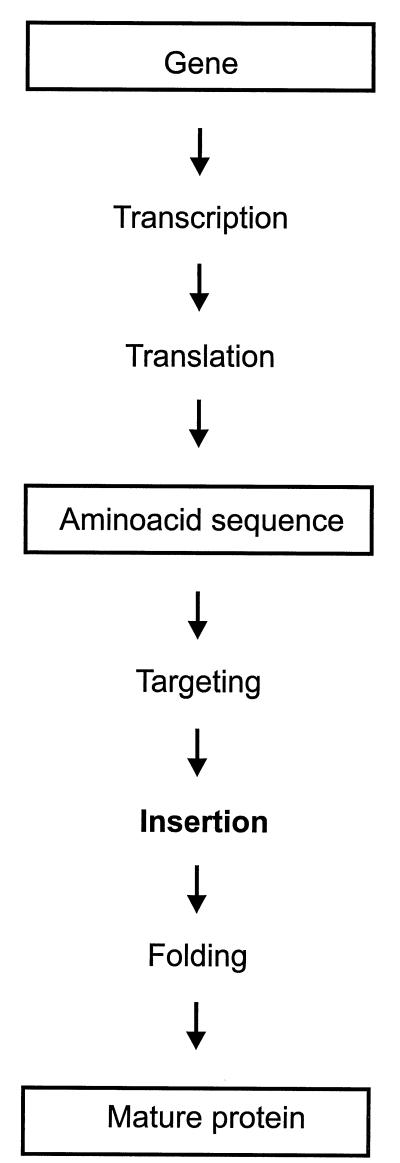
Integral membrane proteins are found in all cellular membranes and carry out many of the functions that are essential to life. The membrane-embedded domains of integral membrane proteins are structurally quite simple, allowing the use of various prediction methods and biochemical methods to obtain structural information about membrane proteins. A critical step in the biosynthetic pathway leading to the folded protein in the membrane is its insertion into the lipid bilayer. Understanding of the fundamentals of the insertion and folding processes will significantly improve the methods used to predict the three-dimensional membrane protein structure from the amino acid sequence. In the first part of this review, biochemical approaches to elucidate membrane protein topology are reviewed and evaluated, and in the second part, the use of similar techniques to study membrane protein insertion is discussed. The latter studies search for signals in the polypeptide chain that direct the insertion process. Knowledge of the topogenic signals in the nascent chain of a membrane protein is essential for the evaluation of membrane topology studies.


Figures

FIG. 1
FIG. 1
Membrane protein biogenesis. In many membrane topology studies, the membrane protein is modified by engineering of the structural gene, while the results are analyzed at the level of the folded protein. The structure of a membrane protein can be deduced from its amino acid sequence only if the different steps leading to the structure are understood.
FIG. 2
FIG. 2
Biochemical approaches to determination of membrane protein topology. (A) Insertions. (B) Fusions.
FIG. 3
FIG. 3
ER translocase complex. The luminal OST is responsible for the glycosylation of membrane proteins in the ER. The active site of OST is located ∼14 residues away from the end of a downstream transmembrane segment and ∼12 residues away from an upstream hydrophobic segment.
FIG. 4
FIG. 4
Cysteine accessibility assay. Labeling of a periplasmic (A) and a cytoplasmic (B) cysteine residue in intact cells. The cells are treated with a detectable and membrane-permeant cysteine reagent (Label; from left to right) with or without pretreatment with a membrane-impermeable cysteine reagent (Block). Following the treatment, the protein is purified from the cells and assayed for labeling.
FIG. 5
FIG. 5
Reaction of a thiol with a maleimide.
FIG. 6
FIG. 6
In vivo biotinylation. Shown is the condensation reaction of biotin and a lysine residue, yielding a biotinylated protein. The reaction is catalyzed by the enzyme biotin ligase.
FIG. 7
FIG. 7
Membrane protein insertion into the ER membrane. The SRP recognizes the first transmembrane segment emerging from the ribosome and targets the whole complex to the ER membrane. The ribosome-nascent chain complex is delivered at the translocon, where translation resumes and the protein is inserted into the membrane.
FIG. 8
FIG. 8
Sequential insertion of hydrophobic sequences. Hydrophobic segments insert into the membrane as they emerge from the ribosome. The orientation of the segment is opposite to the orientation of the previous segment. A segment with an Ncyt-Clumen orientation is termed signal anchor (SA), and a segment with an Nlumen-Ccyt orientation is termed stop transfer (ST).
FIG. 9
FIG. 9
M0 and M1 system. The variable sequence is preceded by the N-terminal region excluding (M0) or including (M1) the first transmembrane segment of the α-subunit of the gastric H+,K+ P-type ATPase and followed by the C-terminal region of the β-subunit of the same enzyme to assay for ST and SA activity, respectively (left). Insertion of the variable sequence into the membrane results in glycosylation of the C-terminal region of the β-subunit in the M0 vector and in the lack of glycosylation in the M1 vector (right).
FIG. 10
FIG. 10
Lep system. Vectors based on Leader peptidase of E. coli. The variable sequence replaces the second transmembrane segment, H2, of Lep (top) to measure SA activity or is inserted in the P2 domain to assay for ST activity (bottom). Membrane insertion of the variable sequence results in glycosylation and lack of glycosylation, respectively (right).
FIG. 11
FIG. 11
Prolactin system. Shown are vectors based on the secretory protein prolactin. The variable sequence is inserted between preprolactin and prolactin (top), the H1 domain of leader peptidase and prolactin (middle), and a hydrophilic domain and prolactin (bottom). The assay of the membrane insertion activity of the variable sequence is based on protection of the prolactin domain against proteolytic degradation (right). The scissors represents the protease.
FIG. 12
FIG. 12
Hypothetical folding intermediates. In the folding intermediates (left), two successive transmembrane segments are not inserted in the membrane. The hydrophobic segment, indicated as a gray box, drives the insertion of the preceding (A and B) or following (C) segment to yield the membrane topologies indicated on the right. The intermediates are based on observations made with the anion exchanger Band 3 (A), the protein translocation complex subunit Sec61 (A and B), and the citrate transporter CitS (C).

Similar articles

Cited by

References

    1. Akabas M H, Karlin A. Identification of acetylcholine receptor channel-lining residues in the M1 segment of the alpha-subunit. Biochemistry. 1995;34:12496–12500. - PubMed
    1. Akabas M H, Kaufmann C, Archdeacon P, Karlin A. Identification of acetylcholine receptor channel-lining residues in the entire M2 segment of the alpha subunit. Neuron. 1994;13:919–927. - PubMed
    1. Akabas M H, Kaufmann C, Cook T A, Archdeacon P. Amino acid residues lining the chloride channel of the cystic fibrosis transmembrane conductance regulator. J Biol Chem. 1994;269:14865–14868. - PubMed
    1. Akabas M H, Stauffer D A, Xu M, Karlin A. Acetylcholine receptor channel structure probed in cysteine-substitution mutants. Science. 1992;258:307–310. - PubMed
    1. Akiyama Y, Ito K. Folding and assembly of bacterial alkaline phosphatase in vitro and in vivo. J Biol Chem. 1993;268:8146–8150. - PubMed

LinkOut - more resources