Review
. 2000 Mar;64(1):51-68.
doi: 10.1128/MMBR.64.1.51-68.2000.

Hepatitis B virus biology

Affiliations
Review

Hepatitis B virus biology

C Seeger et al. Microbiol Mol Biol Rev. 2000 Mar.

Abstract

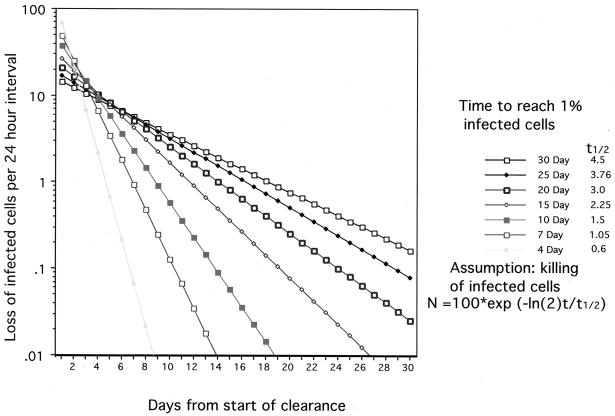
Hepadnaviruses (hepatitis B viruses) cause transient and chronic infections of the liver. Transient infections run a course of several months, and chronic infections are often lifelong. Chronic infections can lead to liver failure with cirrhosis and hepatocellular carcinoma. The replication strategy of these viruses has been described in great detail, but virus-host interactions leading to acute and chronic disease are still poorly understood. Studies on how the virus evades the immune response to cause prolonged transient infections with high-titer viremia and lifelong infections with an ongoing inflammation of the liver are still at an early stage, and the role of the virus in liver cancer is still elusive. The state of knowledge in this very active field is therefore reviewed with an emphasis on past accomplishments as well as goals for the future.


Figures

FIG. 1
FIG. 1
Anatomy of the liver lobule. The major cell types within the liver are illustrated. Blood enters at the portal tracts (encompassing the portal vein, hepatic artery, and bile ductules), distributes laterally through smaller veins to the sinusoidal spaces, and flows toward the central vein. The sinusoidal spaces are lined with endothelial cells and fixed macrophages (Kupffer cells). The endothelial cells are fenestrated, allowing free diffusion between the blood and hepatocytes.
FIG. 2
FIG. 2
Hepadnavirus life cycle. The details of the replication cycle are discussed in the text. Briefly, during initiation of infection the viral rcDNA (or linear DNA) genome, with a protein (the viral reverse transcriptase) attached to the 5′ end of the minus strand and a short RNA attached to the 5′ end of the plus strand, is converted into cccDNA. During this process, both the protein and the RNA are removed. The cccDNA serves as the template for transcription of viral mRNAs (see Fig. 4). One of these, the pregenome, serves as the mRNA for the synthesis of core protein (nucleocapsid subunit) and the viral reverse transcriptase. The reverse transcriptase binds to the 5′ end of its own mRNA template, and the complex is then packaged into nucleocapsids, where viral DNA synthesis occurs (see Fig. 3). Once partially double-stranded DNA has been produced, nucleocapsids can undergo a maturation event that facilitates their acquisition of an outer envelope via budding into the ER. These nucleocapsids can also migrate to the nucleus to increase the copy number of cccDNA. Since cccDNA does not undergo semiconservative replication, all cccDNA is derived from viral DNA made in the cytoplasm via the reverse transcription pathway (217). Accumulation of viral envelope proteins prevents excessive cccDNA formation, which can be toxic to hepatocytes (200, 201).
FIG. 3
FIG. 3
Hepadnavirus DNA replication. (A) Virus particles contain predominantly rcDNA with a complete minus strand and a partially synthesized plus strand. A small amount of linear DNA, formed as a result of aberrant plus-strand priming (in situ priming) is also found in the virus. (B) During initiation of infection, both forms of virion DNA are converted to a cccDNA; however, conversion of the linear form involves illegitimate recombination, which can lead to subsequent defects in the ability of the virus to replicate. cccDNA serves as template for the transcription of the pregenome. The reverse transcriptase binds to the epsilon stem-loop structure near the 5′ end of its own mRNA to facilitate packaging into nucleocapsids and initiation of reverse transcription by a protein-priming mechanism, utilizing a tyrosine located near the amino terminus of the reverse transcriptase itself. (C) Following the synthesis of 4 bases, the polymerase translocates to the 3′ end of the RNA template, where the 4 bases can anneal with complementary sequences. During the elongation of the minus strand to the 5′ end of the pregenome, all but the 5′ 17 to 18 bases of the pregenome, including the CAP and DR1, are degraded by the viral RNase H. (D) The remaining fragment then serves as the primer for plus-strand synthesis, usually following its translocation to DR2, with which it can hybridize because of the sequence identity between DR1 and DR2. A third translocation occurs when the plus strand reaches the 5′ end of the minus strand, to circularize the molecule and allow continued plus-strand elongation. This translocation may be facilitated by the short (∼9-base) terminal redundancy on the minus strand. The plus strand is not completed prior to virion release; a repair reaction to produce a fully double stranded DNA occurs during initiation of a subsequent round of infection. (E) A fraction of the virions have linear genomes because priming of plus-strand DNA synthesis occurs in the absence of primer translocation.
FIG. 4
FIG. 4
Transcriptional and translational map of HBV. The figure shows the physical map of the HBV genome. The inner circle depicts the rcDNA with the reverse transcriptase attached to the 5′ end of the complete minus-strand DNA (solid sphere) and a capped RNA oligomer attached to the 5′ end of the incomplete plus-strand DNA (solid half sphere). The positions of the direct repeats, DR1 and DR2, as well as the positions of the two enhancers, EN1 and EN2, are indicated. The outer circle depicts the three major viral RNAs, the core (C) or pgRNA, the pre-S (L) mRNA, and the S mRNA. The common 3′ ends of the three mRNAs are indicated by the letters A. Not shown in the figure is the putative X mRNA that spans the X coding region and terminates at the site indicated for the other three mRNAs. The four protein-coding regions are shown between the inner and outer circles. They include the precore (PC) and core genes, the polymerase gene, and the X gene. The envelope genes pre-S1 (L), pre-S2 (M), and surface (S) overlap with the polymerase open reading frame.
FIG. 5
FIG. 5
CTL killing and hepatocyte replacement. The kinetics of clearance of infected hepatocytes have been calculated by assuming that CTL killing is a first-order reaction. Each point indicates the fraction of the initial number of infected hepatocytes that were killed in the preceding 24-h period. The different curves were determined using t1/2 values for killing that would produce 99% loss of infected hepatocytes in the indicated times.

References

    1. Albritton L M, Tseng L, Scadden D, Cunningham J M. A putative murine ecotropic retrovirus receptor gene encodes a multiple membrane-spanning protein and confers susceptibility to virus infection. Cell. 1989;57:659–666. - PubMed
    1. Aldrich C E, Coates L, Wu T T, Newbold J, Tennant B C, Summers J, Seeger C, Mason W S. In vitro infection of woodchuck hepatocytes with woodchuck hepatitis virus and ground squirrel hepatitis virus. Virology. 1989;172:247–252. - PubMed
    1. Allen M I, Deslauriers M, Andrews C W, Tipples G A, Walters K A, Tyrrell D L, Brown N, Condreay L D. Identification and characterization of mutations in hepatitis B virus resistant to lamivudine. Lamivudine Clinical Investigation Group. Hepatology. 1998;27:1670–1677. - PubMed
    1. Balsano C, Avantaggiati M L, Natoli G, De M E, Will H, Perricaudet M, Levrero M. Full-length and truncated versions of the hepatitis B virus (HBV) X protein (pX) transactivate the cmyc protooncogene at the transcriptional level. Biochem Biophys Res Commun. 1991;176:985–992. - PubMed
    1. Barker L F, Chisari F V, McGrath P P, Dalgard D W, Kirschstein R L, Almeida J D, Edgington T S, Sharp D G, Peterson M R. Transmission of type B viral hepatitis to chimpanzees. J Infect Dis. 1973;127:648–652. - PubMed

Publication types