Review
. 2000 Mar;64(1):138-52.
doi: 10.1128/MMBR.64.1.138-152.2000.

Higher levels of organization in the interphase nucleus of cycling and differentiated cells

Affiliations
Review

Higher levels of organization in the interphase nucleus of cycling and differentiated cells

A R Leitch. Microbiol Mol Biol Rev. 2000 Mar.

Abstract

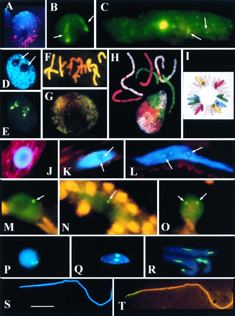
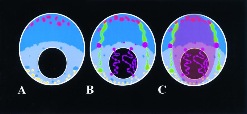
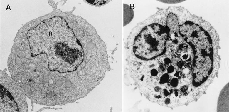
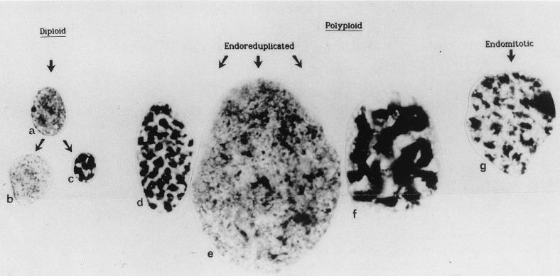
The review examines the structured organization of interphase nuclei using a range of examples from the plants, animals, and fungi. Nuclear organization is shown to be an important phenomenon in cell differentiation and development. The review commences by examining nuclei in dividing cells and shows that the organization patterns can be dynamic within the time frame of the cell cycle. When cells stop dividing, derived differentiated cells often show quite different nuclear organizations. The developmental fate of nuclei is divided into three categories. (i) The first includes nuclei that undergo one of several forms of polyploidy and can themselves change in structure during the course of development. Possible function roles of polyploidy is given. (ii) The second is nuclear reorganization without polyploidy, where nuclei reorganize their structure to form novel arrangements of proteins and chromosomes. (iii) The third is nuclear disintegration linked to programmed cell death. The role of the nucleus in this process is described. The review demonstrates that recent methods to probe nuclei for nucleic acids and proteins, as well as to examine their intranuclear distribution in vivo, has revealed much about nuclear structure. It is clear that nuclear organization can influence or be influenced by cell activity and development. However, the full functional role of many of the observed phenomena has still to be fully realized.


Figures

FIG. 1
FIG. 1
(A and B) Triticum aestivum (wheat) root tip meristematic nuclei. (A) CCS1 labelling for centromeres (digoxigenin-fluorescein isothiocyanate [FITC], cyan fluorescence) at one pole and the telomere consensus sequence (TTTAGGG)n, biotin-Cy3, red fluorescence) at the other. The red dots apparently outside the central nucleus correspond to signal from the telomeric pole of an adjacent nucleus. The nucleus is counterstained for DNA (DAPI stain, blue fluorescence). Photo courtesy of L. Aragon-Alacaide and G. Moore. (B) T. aestivum cv. Beaver carrying two 1Bl/1Rs chromosome arms has 1Rs detected by GISH (digoxigenin-labelled total Secale cereale [rye] DNA-FITC, green fluorescence). Note that the two elongate 1Rs chromosome arm domains (arrows) with the condensed subtelomeric heterochromatin fluoresce more strongly than the remainder of the arm. (C) Protophloem nucleus of T. aestivum cv. Beaver labelled by GISH with total rye DNA (digoxigenin-FITC, green fluorescence). Note that the nucleus is much larger and more elongated than in panel B and is endoreduplicated; the single large 1Rs domain is in the center of the nucleus; there is no evidence of elongate chromosome domains; and fragments of rye signal across the whole volume of the nucleus (arrows). (D and E) Sectioned nucleus from a wheat meristematic cell. (D) The nucleus is counterstained with DAPI (blue fluorescence) for DNA. Note the two spherical nucleoli (arrows). (E) The same nucleus section labelled for rDNA (digoxigenin-labelled pTa71 [59]-FITC, yellow fluorescence). The rDNA signal occurs outside the nucleolus (arrowed in panel D) on a condensed chromatin fiber and inside the nucleolus on chromatin fibers with different levels of condensation (compare with panel D). (F and G) Root tip meristematic metaphase (F) and interphase (G) of the hybrid Hordeum vulgare (barley) × Secale africanum (wild rye) labelled by GISH with total DNA from the wild rye parent (digoxigenin-FITC, yellow fluorescence) and counterstained with propidium iodide for DNA (orange fluorescence). (F) The metaphase plate shows genome separation, with seven chromosomes of wild-rye origin at the periphery and the seven chromosomes of barley origin at the center. (G) Genome separation at interphase with wild-rye chromatin outside the central barley genome. Panels F and G are taken from reference . (H) Chromosome painting of a metaphase and interphase nucleus of female fibroblasts of Muntiacus muntjac vaginalis (Indian munjac), chromosome 1 (biotin-Cy5, white), chromosome 2 (FITC–12-dUTP, green), chromosome X +3 (Cy3-dUTP, red) (157). Note that the chromosomes occur in discrete unpaired domains at interphase. Photograph courtesy of F. Yang and M. Ferguson-Smith. Taken from Chromosome Research. (I) Diagram of human fibroblast prometaphase showing the positions of chromosomes 7, 8, 16, and X (from reference 120). Note that a complete set of identified chromosomes are drawn on each side of the prometaphase, suggesting genome separation. Nagele et al. (120) also suggest that there is an order of chromosomes in each genome (i.e. 7, 16, X, 8) in two antiparallel sets. (J to O) Funaria hygrometrica (moss) nuclei from caulonemata (J to N) and from a thallus cell (O). (J to L) DAPI-stained nuclei (blue fluorescence) from an apical cell (J), cell 8 (K), and cell 15 (L) of the caulonemata filament. Note the increasing size and elongation of nucleus and the accumulation of rDNA heterochromatin (arrows). (M to O) Immunocytochemistry to detect D-polypeptide of the spliceosome complex (FITC detection, green fluorescence). In addition to a uniform dispersal of signal across the nucleus but outside the nucleolus, there is one coiled body in the nuclei of cells 4 (M) and 10 (N) of caulonema and two coiled bodies in the nuclei of thallus cells (arrows). All coiled bodies are associated with the nucleolus (O). (P to T) Spermatogenesis in Schistocerca gregaria (locust) stained blue for DNA with DAPI (blue fluorescence) and labelled for rDNA (pTa71, digoxigenin-FITC, green/cyan fluorescence). (P to R) Detection of rDNA in double exposures with DAPI (blue/cyan fluorescence). (P) Early spermatid nucleus with two rDNA loci. (Q and R) Increasingly mature and elongated spermatid nuclei. Note the elongating rDNA loci. (S and T) Fully mature and elongated spermatozoan nucleus, DAPI stained (S) and probed for rDNA (T, yellow fluorescence). Note that all the rDNA signal (yellow) is basal to the nucleus, suggesting intranuclear migration of rDNA. Scale bars, 15 μm for panels A, B, C, F, G, M, N, O, P, Q, R, S, and T; 10 μm for panels D and E; and 20 μm for panels H, J, K, and L.
FIG. 2
FIG. 2
Diagrammatic projections of a root tip meristematic nucleus of a wheat cultivar that carries a 1Bl/1Rs translocation. The intensity of the blue coloration gives an indication of overall DNA condensation levels across the nucleus. The black circle is a nucleolus. (A) The nucleus is drawn in the Rabl configuration, with centromeres clustered at one pole (red) and telomeres at the other pole (yellow). (B) The same nucleus with the inclusion of the 1Rs chromosome arm domains (green), showing different levels of condensation. The rDNA loci (purple) on the 1Rs chromosome arms are drawn condensed and inactive and occupy the region of the nucleus with the highest overall level of DNA condensation. The rDNA loci from wheat chromosomes 6B and 5D are drawn extended through the nucleolus and with varying thickness to illustrate different levels of condensation and activity along each locus. (C) The same nucleus with the inclusion of a central domain to illustrate the possibility of genome separation within these nuclei.
FIG. 3
FIG. 3
Electron micrograph of a stimulated lymphocyte (A) and a human granulocyte (B). (A) The nucleus (n) has many decondensed chromatin fibers throughout its volume. Condensed fibers are found at the nuclear periphery. (B) The highly lobed nucleus has a thin filament of chromatin connecting the lobes (arrow). Large amounts of condensed chromatin are found close to the nuclear envelope and in clumps internal to the nucleus. Magnification, ×5,000.
FIG. 4
FIG. 4
(a to e and g) Variation in the morphology and ploidy levels interphase nuclei from human female cell types. (f) Nucleus from a pea root. Taken from reference .
FIG. 5
FIG. 5
Graph to show the association of homologous chromosomes in nondividing adult human Sertoli cells (open) and stimulated lymphocytes (solid). The distribution of chromosomes is significantly different between Sertoli cells and stimulated lymphocytes for chromosomes 3, 7, 8, and 17. Only for the acrocentric chromosomes 13 and 21, which form nucleoli in the center of the nucleus in both cell types, are there similar distributions. Chandley et al. (37) suggested that homologue pairing was a feature associated with cell differentiation.

Similar articles

Cited by

References

    1. Abney J R, Cutler B, Fillbacj M L, Axelrod D, Scalettar B A. Chromatin dynamics in interphase nuclei and its implications for nuclear structure. J Cell Biol. 1997;137:1459–1468. - PMC - PubMed
    1. Abranches R, Bevan A F, Aragon-Alcaide L, Shaw P J. Transcription sites are not correlated with chromosome territories in wheat nuclei. J Cell Biol. 1998;143:5–12. - PMC - PubMed
    1. Anamthawat-Jönsson K, Heslop-Harrison J S. Centromeres, telomeres and chromatin in the interphase nucleus of cereals. Caryologia. 1990;43:205–213.
    1. Appels R, Honeycutt R L. rDNA: evolution over a billion years. In: Datta S K, editor. DNA systematics. II. Plants. Boca Raton, Fla: CRC Press Inc.; 1986. pp. 81–135.
    1. Aragon-Alcaide L, Miller T, Schwarzacher T, Reader S, Moore G. A cereal centromeric sequence. Chromosoma. 1996;105:261–268. - PubMed

Publication types

LinkOut - more resources