Comparative Study
. 2000 Mar 15;20(6):2112-20.
doi: 10.1523/JNEUROSCI.20-06-02112.2000.

Axonal transport of microtubule-associated protein 1B (MAP1B) in the sciatic nerve of adult rat: distinct transport rates of different isoforms

Affiliations
Comparative Study

Axonal transport of microtubule-associated protein 1B (MAP1B) in the sciatic nerve of adult rat: distinct transport rates of different isoforms

D Ma et al. J Neurosci. .

Abstract

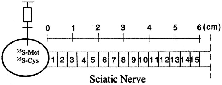
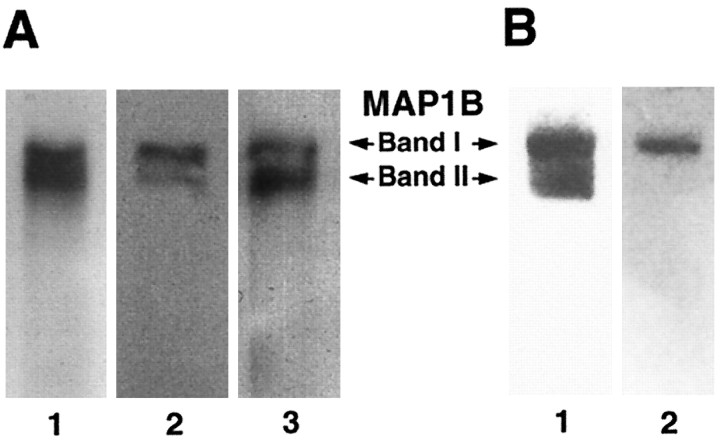
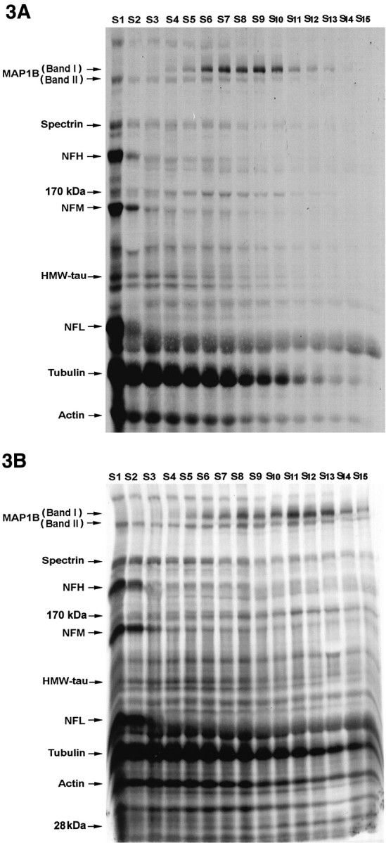
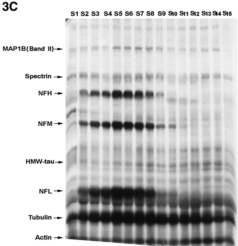
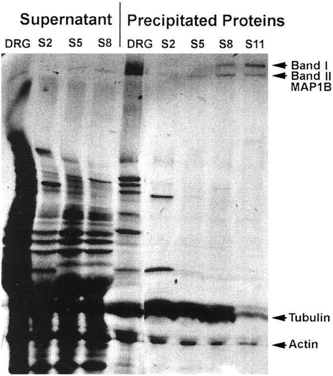
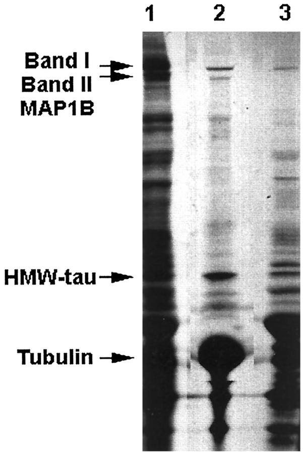
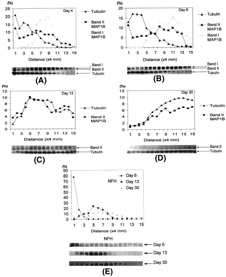
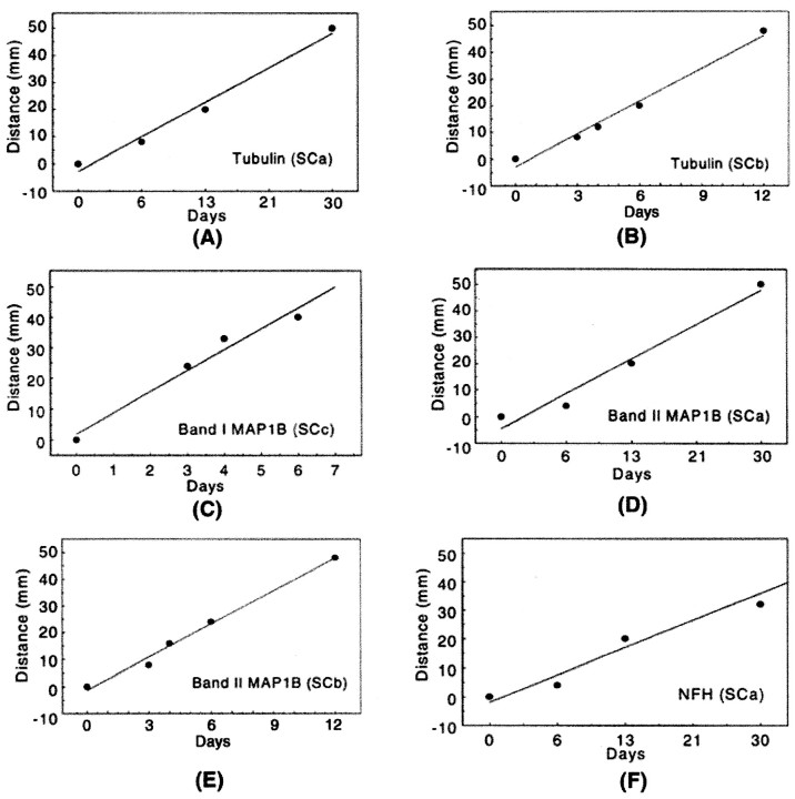
Cytoskeletal proteins are axonally transported with slow components a and b (SCa and SCb). In peripheral nerves, the transport velocity of SCa, which includes neurofilaments and tubulin, is 1-2 mm/d, whereas SCb, which includes actin, tubulin, and numerous soluble proteins, moves as a heterogeneous wave at 2-4 mm/d. We have shown that two isoforms of microtubule-associated protein 1B (MAP1B), which can be separated on SDS polyacrylamide gels on the basis of differences in their phosphorylation states (band I and band II), were transported at two different rates. All of band I MAP1B moved as a coherent wave at a velocity of 7-9 mm/d, distinct from slow axonal transport components SCa and SCb. Several other proteins were detected within the component that moved at the velocity of 7-9 mm/d, including the leading wave of tubulin and actin. The properties of this component define a distinct fraction of the slow axonal transport that we suggest to term slow component c (SCc). The relatively fast transport of the phosphorylated MAP1B isoform at 7-9 mm/d may account for the high concentration of phosphorylated MAP1B in the distal end of growing axons. In contrast to band I MAP1B, the transport profile of band II was complex and contained components moving with SCa and SCb and a leading edge at SCc. Thus, MAP1B isoforms in different phosphorylation states move with distinct components of slow axonal transport, possibly because of differences in their abilities to associate with other proteins.


Figures

Fig. 1.
Fig. 1.
Diagram showing the injection of the [35S]methionine and [35S]cysteine mixture into the DRG and the orientation of the sciatic nerve segments used for fluorographic studies.
Fig. 2.
Fig. 2.
Western blots probed with the MAP1B polyclonal antibody 1BNR (1:5000 dilution). A, Heterogeneity of the MAP1B protein in rat brain at postnatal day 7 (lane 1; 20 μg), adult DRG (lane 2; 30 μg), and sciatic nerve (lane 3; 40 μg) is shown. B, In rat brain the antibody 1BNR detected both bands I and II (lane 1), but the monoclonal 1BP antibody, which recognizes a phosphorylated isoform of MAP1B, reacted specifically with band I (lane 2).
Fig. 3.
Fig. 3.
Fluorographs illustrating the slow transport profiles of radiolabeled proteins in sciatic axons of DRG neurons by 4 d (A), 6 d (B), and 13 d (C) after injection of [35S]methionine and [35S]cysteine. The lanes(S1–S15) in these gels represent 15 consecutive 4 mm sciatic nerve segments progressing distally from the DRG.HMW-tau, High-molecular weight tau (110 kDa);MAP1B, microtubule-associated protein 1B (320–340 kDa);NFH, high-molecular weight neurofilament (200 kDa);NFL, low-molecular weight neurofilament (70 kDa);NFM, middle-molecular weight neurofilament (160 kDa);S1–S15, segments 1–15.
Fig. 3.
Fig. 3.
Fluorographs illustrating the slow transport profiles of radiolabeled proteins in sciatic axons of DRG neurons by 4 d (A), 6 d (B), and 13 d (C) after injection of [35S]methionine and [35S]cysteine. The lanes(S1–S15) in these gels represent 15 consecutive 4 mm sciatic nerve segments progressing distally from the DRG.HMW-tau, High-molecular weight tau (110 kDa);MAP1B, microtubule-associated protein 1B (320–340 kDa);NFH, high-molecular weight neurofilament (200 kDa);NFL, low-molecular weight neurofilament (70 kDa);NFM, middle-molecular weight neurofilament (160 kDa);S1–S15, segments 1–15.
Fig. 4.
Fig. 4.
Immunoprecipitation identifying the radiolabeled MAP1B isoforms in the L5 DRG and sciatic nerve segments 2, 5, 8, and 11 by day 6 after injection. Total labeled protein homogenates were incubated with the specific MAP1B polyclonal antibody 1BNR and precipitated by protein A-bound agarose beads. DRG, Dorsal root ganglion; MAP1B, microtubule-associated protein 1B (320–340 kDa); S2, segment 2;S5, segment 5; S8, segment 8;S11, segment 11.
Fig. 5.
Fig. 5.
Taxol preparations of microtubules showing radiolabeled MAP1B proteins through in vitro microtubule assembly from the sciatic nerve segments 8–14 obtained 6 d after injection. The taxol-assembled microtubule pellet (lane 2) contains both band I and band II MAP1B isoforms. Total radiolabeled proteins in the supernatant extracted by homogenization buffer containing SDS are shown in lane 1, whereas the nonmicrotubule supernatant is shown in lane 3.HMW-tau, High-molecular weight tau (110 kDa);MAP1B, microtubule-associated protein 1B.
Fig. 6.
Fig. 6.
The distribution of radiolabeled tubulin and MAP1B in the sciatic nerve axons at 4 d (A), 6 d (B), 13 d (C), and 30 d (D) after injection and the distribution of radiolabeled NFH in the sciatic nerve axons at 6, 13, and 30 d after injection (E). The amount of radioactivity in consecutive 4 mm segments of the sciatic nerve, expressed relative to the total radioactivity of each specific protein in the sciatic nerve, is plotted against distance from the DRG. The data shown are the average of three to four independent determinations. NFH, High-molecular weight neurofilament; MAP1B, microtubule-associated protein 1B.
Fig. 7.
Fig. 7.
Regression analysis of three slow components of axonal transport in adult rat sciatic nerves. Tubulin is transported with both SCa (A) and SCb (B), whereas neurofilament proteins including NFH are exclusively transported with SCa (F). Interestingly, the band I MAP1B isoform and a fast wave of the band II MAP1B isoform move with SCc (C), whereas the band II MAP1B isoform moves mainly with SCa (D) and SCb (E). MAP1B, Microtubule-associated protein 1B; NFH, high-molecular weight neurofilament; SC, slow component.

Similar articles

Cited by

References

    1. Baas PW, Yu W. A composite model for establishing the microtubule arrays of the neuron. Mol Neurobiol. 1996;12:145–161. - PubMed
    1. Baitinger C, Willard M. Axonal transport of synapsin I-like proteins in rabbit retinal ganglion cells. J Neurosci. 1987;7:3723–3735. - PMC - PubMed
    1. Bamburg JR, Bray D, Chapman K. Assembly of microtubules at the tip of growing axons. Nature. 1986;321:788–790. - PubMed
    1. Black MM, Lasek RJ. A difference between the proteins conveyed in the fast component of axonal transport in guinea pig hypoglossal and vagus motor neurons. J Neurobiol. 1978;9:433–443. - PubMed
    1. Black MM, Lasek RJ. Axonal transport of actin: slow component b is the principal source of actin for the axon. Brain Res. 1979;171:401–413. - PubMed

Publication types

MeSH terms