. 2000 Mar 15;20(6):2121-30.
doi: 10.1523/JNEUROSCI.20-06-02121.2000.

Presynaptic P2X receptors facilitate inhibitory GABAergic transmission between cultured rat spinal cord dorsal horn neurons

Affiliations

Presynaptic P2X receptors facilitate inhibitory GABAergic transmission between cultured rat spinal cord dorsal horn neurons

S Hugel et al. J Neurosci. .

Abstract

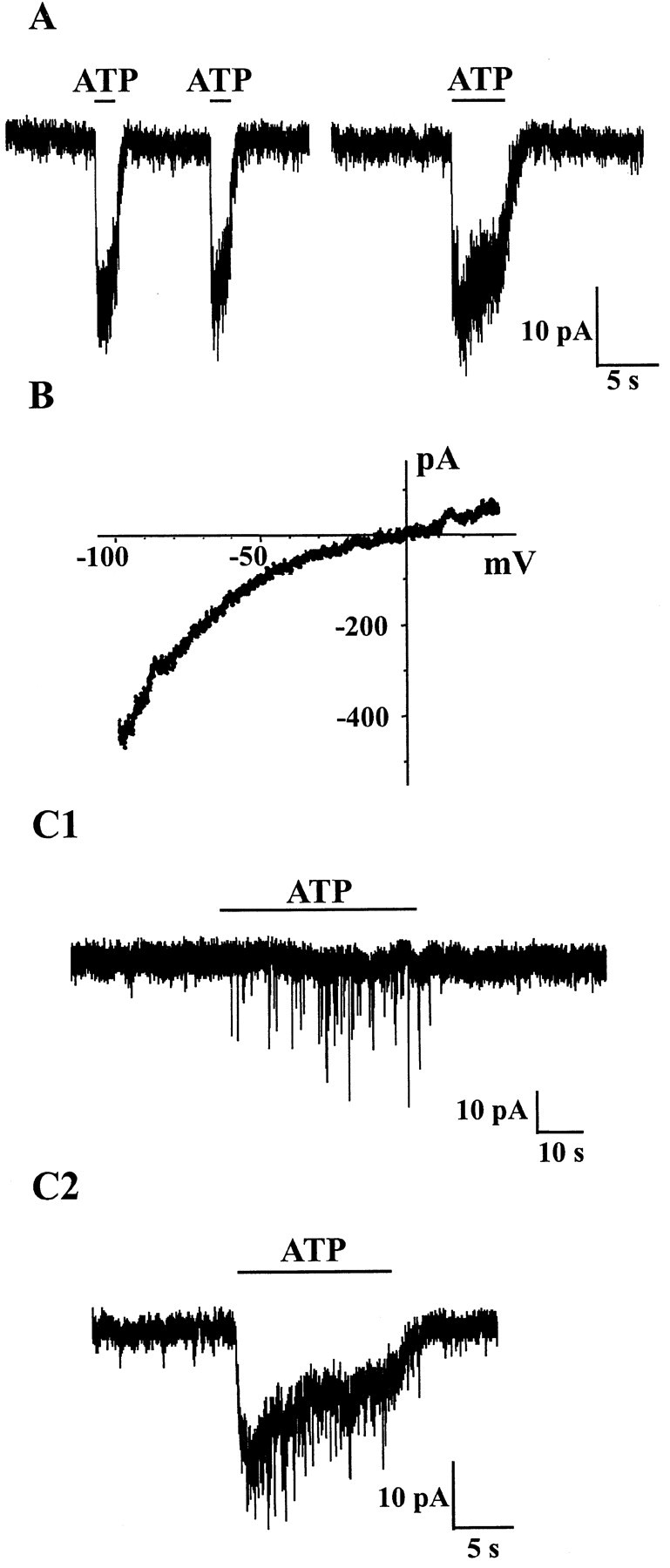
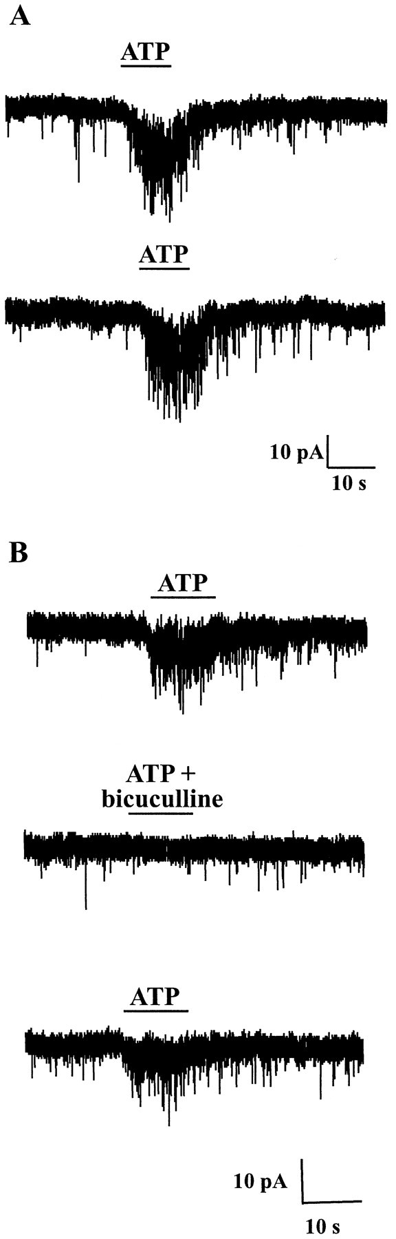
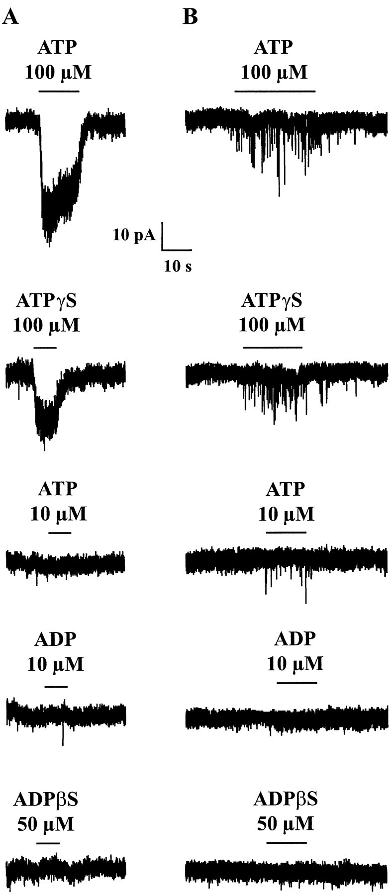
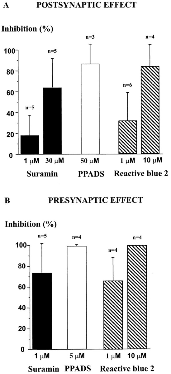
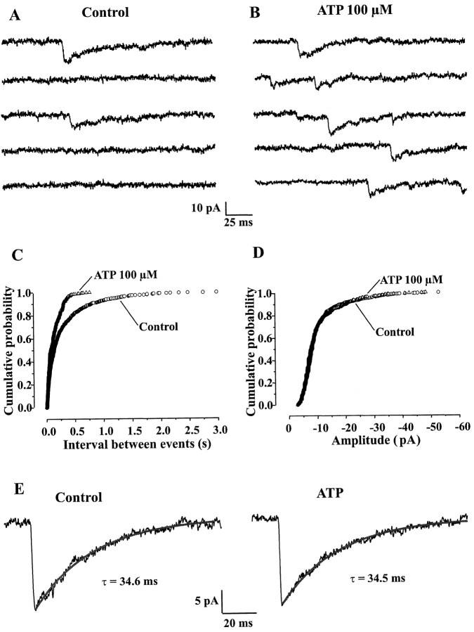
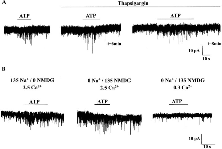
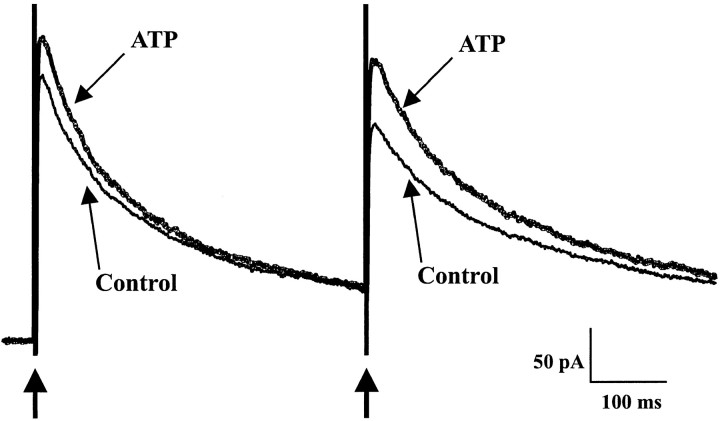
The superficial layers of the spinal cord dorsal horn (DH) express P2X2, P2X4, and P2X6 subunits entering into the formation of ionotropic (P2X) receptors for ATP. Using a culture system of laminae I-III from neonatal rat DH, we show that ATP induced a fast nonselective cation current in 38% of the neurons (postsynaptic effect). ATP also increased the frequency of miniature IPSCs (mIPSCs) mediated by GABA(A) receptors or by glycine receptors in 22 and 9%, respectively, of the neurons tested (presynaptic effect) but had no effect on glutamatergic transmission. The presynaptic effect of ATP on GABAergic transmission was not significantly affected by thapsigargin (1 microM) but was completely dependent on Ca(2+) influx. Presynaptic and postsynaptic effects were inhibited by suramin, pyridoxalphosphate-6-azophenyl-2',4'-disulfonic acid, and reactive blue and were not reproduced by uridine 5'-triphosphate (UTP) or adenosine 5'-O-(2-thiodiphosphate) (ADP-beta-S), suggesting the implication of ionotropic P2X rather than of metabotropic P2Y receptors. alphabeta-methylene-ATP (100 microM) did not reproduce the effects of ATP. ATP reversibly increased the amplitude of electrically evoked GABAergic IPSCs and reduced paired-pulse inhibition or facilitation without affecting IPSC kinetics. This effect was preferentially, but not exclusively, observed in neurons coreleasing ATP and GABA. We conclude that in cultured DH neurons, the effects of ATP are mediated by P2X receptors having a pharmacological profile dominated by the P2X2 subunit. The presynaptic receptors might underlie a modulatory action of ATP on a subset of GABAergic interneurons involved in the spinal processing of nociceptive information.


Figures

Fig. 1.
Fig. 1.
Presynaptic and postsynaptic effects of ATP in cultured DH neurons. In this and all other figures, recordings were made in the presence of CNQX (10 μm), d-APV (30 μm), and strychnine (1 μm) in the extracellular medium to block fast glutamatergic and glycinergic synaptic transmissions. TTX (0.5 μm) was also present to record GABAergic mIPSCs. A, Local applications of ATP induced fast inward currents in the postsynaptic neuron. Two successive and close (10 sec interval) ATP (100 μm) applications elicited currents of similar amplitudes. During a long-lasting (5 sec) application of ATP, the current decayed only partially and slowly, indicating the absence of significant desensitization.B, Current–voltage (I–V) relationship of the ATP (100 μm)-induced current obtained by applying a 2 sec lasting monotonic voltage ramp from −100 mV to +30 mV. The reversal potential was close to 0 mV, and theIV relationship showed marked inward rectification at negative membrane potentials. C1, Application of ATP (100 μm) reversibly increased the frequency of mIPSCs recorded as fast downward deflections of the current trace. C2, Coexistence of presynaptic and postsynaptic effects of ATP. In some cells, ATP (100 μm) induced both an inward current and an increase in mIPSC frequency. All traces were recorded at an HP of −60 mV. In A,C1, and C2, the horizontal black bars represent the duration of ATP application.
Fig. 2.
Fig. 2.
ATP increased the frequency of GABAergic mIPSCs in a reproducible manner. A, Two successive ATP (100 μm) applications spaced by a 2 min interval induced a similar (reproducible) increase in mIPSC frequency. B, When coapplied with bicuculline (10 μm), a selective antagonist of GABAA receptors, ATP (100 μm) no longer increased the frequency of synaptic events, indicating that ATP increased the frequency of GABAA receptor-mediated mIPSCs. The inward current developing slowly during the application of ATP was caused by the temporal summation of mIPSCs but not by a direct activation of postsynaptic ATP receptors, as demonstrated by its blockade by bicuculline. HP, −60 mV.
Fig. 3.
Fig. 3.
Effects of P2X and P2Y receptor agonists on cultured DH neurons displaying a postsynaptic (A) or presynaptic (B) response. A, ATP (100 μm) induced an inward current that was partially reproduced by the same concentration of ATP-γ-S, a nonhydrolyzable analog of ATP. ATP (10 μm), ADP (10 μm), or ADP-β-S (50 μm), an agonist at P2Y1 receptors, had no effect in the same cell. B, ATP (100 μm) increased the frequency of GABAergic mIPSCs. This effect was mimicked by ATP-γ-S (100 μm) and to a lesser extent by a lower concentration of ATP (10 μm) or ADP (10 μm). ADP-β-S (50 μm) was without effect. For each panel (column), all recordings were from the same cell. HP, −60 mV.
Fig. 4.
Fig. 4.
Effects of P2X receptor antagonists on postsynaptic and presynaptic actions of ATP. Different concentrations of suramin (black bars), PPADS (white bar), and reactive blue 2 (hatched bars) were tested on postsynaptic (A) and presynaptic (B) responses to ATP (100 μm). The histogram represents the percentage of inhibition by each antagonist of the control response to ATP, which was determined as the amplitude of the membrane current (POSTSYNAPTIC EFFECT) or the increase in frequency of mIPSCs (PRESYNAPTIC EFFECT) induced by ATP (100 μm) in the absence of antagonists. Error bars represent SEM. HP, −60 mV.
Fig. 5.
Fig. 5.
ATP increased the frequency but did not alter the amplitude or the kinetic characteristics of GABAergic mIPSCs.A, B, In a neuron displaying mIPSCs under control conditions (A), application of ATP (100 μm) markedly increased the frequency of these synaptic currents (B). C, Cumulative probability histogram of time intervals between successive mIPSCs, before and during ATP (100 μm) application. ATP shifted the distribution of interevent intervals to the left, indicating an increase in mIPSC frequency. D, Cumulative probability histogram of mIPSC amplitudes. Both distributions match perfectly and were not significantly different (Kolmogorov–Smirnov test), indicating that ATP did not modify the amplitude of mIPSCs. E, ATP (100 μm) had no effect on mIPSCs decay kinetics. Averaged traces from 10 events recorded under control conditions (left trace) or during application of 100 μm ATP (right trace) are represented. τ is the value of the time constant of the single exponential fit represented as thesolid line superimposed on each trace. All data are from the same neuron. HP, −60 mV.
Fig. 6.
Fig. 6.
ATP-evoked increase in mIPSC frequency was strictly dependent on extracellular Ca2+.A, Under control conditions, ATP (100 μm) induced an increase in the frequency of mIPSCs. During bath perfusion of thapsigargin (1 μm), ATP (100 μm) was applied at 2 min intervals. The third (t = 6 min) and fourth (t = 8 min) applications in thapsigargin are represented. Note that thapsigargin did not block the ATP response.B, ATP (100 μm) was applied in extracellular solutions containing different concentrations of Na+ and Ca2+ ions. Na+ was replaced by NMDG. The numberson top of each trace indicate (in millimolar) the concentrations of Na+, NMDG, and Ca2+ for each experimental condition. ATP was still able to increase mIPSC frequency in the absence of external Na+(middle trace), but this effect was no longer observed when the external Ca2+ concentration was lowered from 2.5 to 0.3 mm (right trace). HP, −60 mV.
Fig. 7.
Fig. 7.
Effect of ATP on GABAergic eIPSCs. A paired-pulse stimulation protocol consisting of two identical electrical pulses (0.1 msec duration) separated by 400 msec was applied at a frequency of 0.1 Hz. Arrows indicate the stimulation artifacts corresponding to the electrical stimuli. In the presence of ATP (100 μm, bold trace), the amplitude of both eIPSCs was increased (potentiation of the first eIPSC, 18%; potentiation of the second eIPSC, 29%), and the paired-pulse ratio was decreased (by 50%), indicating a presynaptic mechanism of action. The effect of ATP was completely reversible (data not shown). The control and wash traces are averages of 20 individual consecutive traces and that in ATP is an average of 10 traces. HP, 0 mV.

References

    1. Bardoni R, Goldstein PA, Lee CJ, Gu JG, MacDermott AB. ATP P2X receptors mediate fast synaptic transmission in the dorsal horn of the rat spinal cord. J Neurosci. 1997;17:5297–5304. - PMC - PubMed
    1. Boehm S. ATP stimulates sympathetic transmitter release via presynaptic P2X purinoceptors. J Neurosci. 1999;19:737–746. - PMC - PubMed
    1. Burnashev N. Calcium permeability of glutamate-gated channels in the central nervous system. Curr Opin Neurobiol. 1996;6:311–317. - PubMed
    1. Burnstock G. The past, present and future of purine nucleotides as signalling molecules. Neuropharmacology. 1997;36:1127–1139. - PubMed
    1. Collo G, North RA, Kawashima E, Merlo-Pich E, Neidhart S, Surprenant A, Buell G. Cloning of P2X5 and P2X6 receptors and the distribution and properties of an extended family of ATP-gated ion channels. J Neurosci. 1996;16:2495–2507. - PMC - PubMed

Publication types

MeSH terms

LinkOut - more resources