. 2000 Mar 15;20(6):2229-37.
doi: 10.1523/JNEUROSCI.20-06-02229.2000.

Distinct roles for ionotropic and metabotropic glutamate receptors in the maturation of excitatory synapses

Affiliations

Distinct roles for ionotropic and metabotropic glutamate receptors in the maturation of excitatory synapses

S N Gomperts et al. J Neurosci. .

Abstract

We used the single-cell culture preparation to study the role of activity in the development of glutamatergic synapses in vitro. Rat hippocampal cells grown in isolation on glial islands formed functional autaptic connections and continued to elaborate new synapses throughout the 2 week investigation, resulting in increases in both the evoked AMPA receptor (AMPAR) and NMDA receptor (NMDAR) components of the EPSC. Synaptogenesis was not prevented by chronic blockade of sodium channels or all of the known glutamate receptors. Analysis of miniature EPSCs revealed that AMPAR quantal size doubled over time in vitro whereas NMDAR quantal size remained constant. However, the proportion of synaptic responses mediated only by NMDARs increased over time in vitro. The increase in AMPAR quantal size was prevented by TTX and ionotropic glutamate receptor antagonists, whereas the increase in the proportion of NMDAR-only synapses was prevented by metabotropic glutamate receptor antagonists. Notably, chronic NMDAR blockade incubation did not block the formation of the AMPAR EPSC, indicating that NMDAR-dependent plasticity is not necessary for the onset of AMPAR synaptic transmission in this system. We conclude that action potentials and ionotropic glutamate receptor activation are necessary for the developmental increase in AMPAR quantal size and that metabotropic glutamate receptor activation is required for the production of NMDAR-only synapses, but none of these is essential for synapse formation.


Figures

Fig. 1.
Fig. 1.
Synaptogenesis is ongoing in autaptic cells.A, The evoked AMPAR EPSC increases with time in vitro. A1,Representative average traces from 4 and 12 div cells of AMPAR EPSCs (isolated with APV) superimposed on the action potential artifact (isolated with APV and NBQX) are shown. Note the different calibrations. Traces are the average of 15 responses in either 100 μm APV or 100 μm APV with 5 μm NBQX.A2, In the subpopulation of cells that show evoked responses, the evoked AMPAR EPSC increases developmentally [n = 33 (3–6 div); n = 6 (7–10 div); n = 36 (11–14 div)].B, The proportion of neurons that have evoked responses increases with time in vitro. Neurons were identified by the presence of action potentials and were tested for AMPAR and NMDAR EPSCs. Data in each age group were pooled by day [n = 4 (2 div); n = 83 (3–6 div); n = 14 (7–10 div); n = 16 (11–14 div)]. C, The frequency of miniature AMPAR EPSCs undergoes a marked developmental increase [n= 4 (2 div); n = 7 (3–6 div);n = 19 (11–14 div)].
Fig. 2.
Fig. 2.
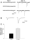
AMPAR quantal size increases over development.A, Representative recordings in 100 μm APV from a day 5 cell and a day 12 cell are shown.B, Representative averaged traces of spontaneous events derived in APV from the day 5 cell (49traces; left) and the day 12 cell (97 traces; right) are shown. C, The mean AMPAR mEPSC amplitude is larger in older cells [n = 8 (3–5 div); n = 18 (11–14 div); p < 0.01].
Fig. 3.
Fig. 3.
NMDAR EPSCs also increase over development.A, The evoked NMDAR EPSC increases developmentally. A1, Representative evoked NMDAR EPSCs from a day 4 cell (left) and a 12 day cell (right) cell demonstrate the large developmental increase in amplitude. Each trace is the average of 10 events. Note the different calibrations.A2, This increase was consistent for the population of cells examined [n = 34 (3–5 div);n = 20 (11–14 div)]. B, The quantal amplitude of the NMDAR component of dual-component mEPSCs is stable over the developmental period. B1, Averaged representative traces from a day 4 (left) and a day 12 (right) cell demonstrate that the sizable increase in AMPAR quantal amplitude is not accompanied by a change in the NMDAR component of mEPSCs. The isolated AMPAR mEPSC, derived in APV, is shown superimposed and scaled to the peak of the dual-component event. One hundred eleven dual-component events and 27 events derived in APV comprise the averagetraces of the young cell. Forty-four dual-component and 38 APV events comprise the average traces of the old cell. B2, Average NMDAR quantal amplitudes from 5 young and 13 old cells are similar. C,The AMPAR/NMDAR amplitude ratio of evoked EPSCs decreases twofold developmentally. AMPAR and NMDAR components were isolated pharmacologically and collected sequentially as the average of 10–30traces each from 37 young and 46 old cells (p < 0.01). D, The fraction of NMDAR-only synapses increases over development in isolated cells. Differences in NMDAR and AMPAR quantal contents calculated from a comparison of evoked and spontaneous dual-component events are used to make this calculation. The results are displayed in terms of a linear function, the r.c.i., which ranges from all NMDAR-only synapses to all AMPAR-only synapses. Although the young cell r.c.i. is not significantly different from zero, the old cell r.c.i. is significantly different from zero (young, n = 5; old,n = 9; young vs old, p < 0.01; old vs zero, p < 0.01).
Fig. 4.
Fig. 4.
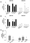
Chronic glutamate receptor blockade reduces both AMPAR quantal size and the fraction of NMDAR-only synapses.A, Evoked AMPAR and NMDAR EPSC amplitudes are unaffected by combined chronic glutamate receptor blockade with APV, NBQX, and MCPG but are differentially affected by APV, NBQX, and MCPG. No manipulation significantly affected AMPAR EPSC amplitude (filledbars). APV significantly increased and MCPG significantly decreased NMDAR EPSC amplitude (openbars) (AMPAR and NMDAR EPSCs, control, n = 18, 20; GluR blockade,n = 13, 12; APV, n= 12, 12; NBQX, n = 9, 9; MCPG,n = 9, 12, respectively; NMDAR EPSCs, APV vs control, *p < 0.05; MCPG vs control, *p < 0.05). B, AMPAR quantal amplitude is reduced by ionotropic but not metabotropic glutamate receptor antagonists, but NMDAR quantal amplitude is unaffected. Glutamate receptor blockade, APV, and NBQX all significantly reduced AMPAR quantal size without affecting NMDAR quantal size (AMPAR and NMDAR mEPSCs, control, n = 18, 13; GluR blockade,n = 8, 8; APV, n = 11, 9; NBQX, n = 6, 6; MCPG, n= 10, 10, respectively; AMPAR mEPSCs, GluR blockade vs control, *p < 0.01; APV vs control, *p< 0.01; NBQX vs control, *p < 0.05).C, The fraction of NMDAR-only synapses, estimated from same-cell comparisons of evoked and spontaneous dual-component events, is reduced by glutamate receptor blockade and MCPG but not by APV or NBQX (control, n = 9; GluR blockade,n = 7; APV, n = 6; NBQX,n = 6; MCPG, n = 9; GluR blockade vs control, *p < 0.05; MCPG vs control, *p < 0.01).
Fig. 5.
Fig. 5.
Chronic metabotropic glutamate receptor blockade reduces the proportion of NMDAR-only puncta. A, Images show immunocytochemical localization of GluR1 (top) and NR1(middle) in 2-week-old autaptic neurons grown in the absence (left) or presence (right) of the metabotropic glutamate receptor antagonist MCPG. Openarrows in overlayed images (bottom) identify colocalized NR1 and GluR1 puncta. Whitearrowheads identify examples of isolated NR1 puncta.B, Cells grown in MCPG had fewer NR1+/GluR1− puncta than did cells grown in normal conditions (p< 0.01; n = 31 control cells, 26 MCPG-treated cells from three dissections).
Fig. 6.
Fig. 6.
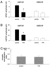
Chronic action potential blockade selectively interferes with the maturational increase in AMPAR quantal size.A, Evoked AMPAR EPSC amplitude but not NMDAR EPSC amplitude is reduced by chronic treatment with TTX.Filledbars represent AMPAR events;openbars represent NMDAR events (AMPAR and NMDAR EPSCs, control, n = 18, 20; TTX,n = 8, 7, respectively; AMPAR EPSCs, TTX vs control, *p < 0.05). B, Mean AMPAR but not NMDAR quantal amplitude is reduced by TTX (AMPAR and NMDAR mEPSCs, control, n = 18, 13; TTX,n = 8, 8, respectively; AMPAR mEPSCs, TTX vs control, *p < 0.01). C, The fraction of NMDAR-only synapses estimated from same-cell comparisons of evoked and spontaneous dual-component events is not affected by TTX (control, n = 9; TTX, n = 5).

Similar articles

Cited by

References

    1. Allen CN, Brady R, Swann J, Hori N, Carpenter DO. N-Methyl-d-aspartate (NMDA) receptors are inactivated by trypsin. Brain Res. 1988;458:147–150. - PubMed
    1. Allison DW, Gelfand VI, Spector I, Craig AM. Role of actin in anchoring postsynaptic receptors in cultured hippocampal neurons: differential attachment of NMDA versus AMPA receptors. J Neurosci. 1998;18:2423–2436. - PMC - PubMed
    1. Basarsky TA, Parpura V, Haydon PG. Hippocampal synaptogenesis in cell culture: developmental time course of synapse formation, calcium influx, and synaptic protein distribution. J Neurosci. 1994;14:6402–6411. - PMC - PubMed
    1. Bekkers JM, Stevens CF. NMDA and non-NMDA receptors are co-localized at individual excitatory synapses in cultured rat hippocampus. Nature. 1989;341:230–233. - PubMed
    1. Benson DL, Cohen PA. Activity-independent segregation of excitatory and inhibitory synaptic terminals in cultured hippocampal neurons. J Neurosci. 1996;16:6424–6432. - PMC - PubMed

Publication types

MeSH terms

LinkOut - more resources