. 2000 Mar 15;20(6):2247-54.
doi: 10.1523/JNEUROSCI.20-06-02247.2000.

Late retinal progenitor cells show intrinsic limitations in the production of cell types and the kinetics of opsin synthesis

Affiliations

Late retinal progenitor cells show intrinsic limitations in the production of cell types and the kinetics of opsin synthesis

M J Belliveau et al. J Neurosci. .

Abstract

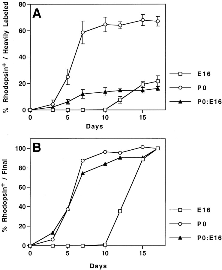
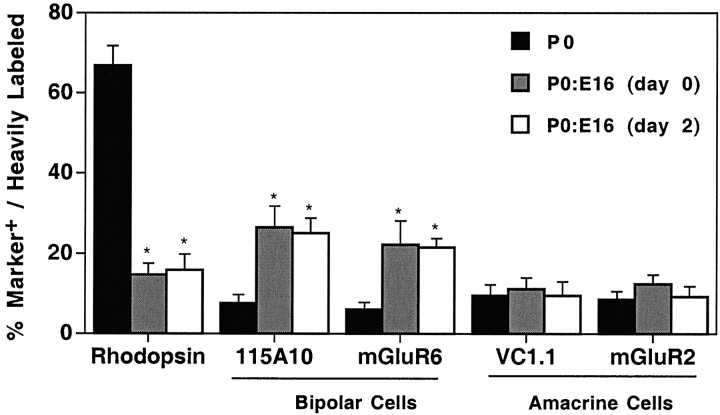
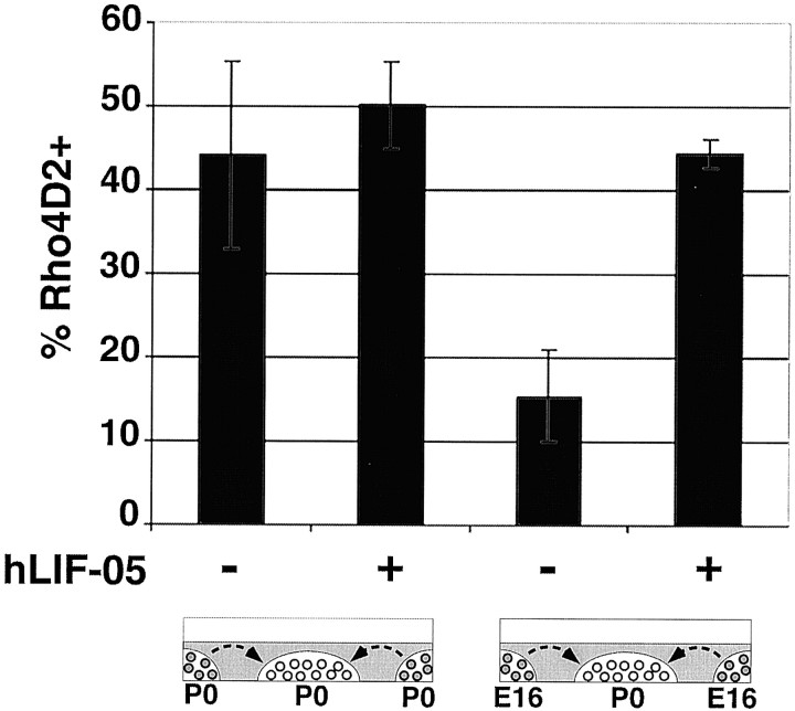
The seven major cell classes of the vertebrate neural retina arise from a pool of multipotent progenitor cells. Several studies suggest a model of retinal development in which both the environment and the progenitor cells themselves change over time (). To test this model, we used a reaggregate culture system in which a labeled population of progenitor cells from the postnatal rat retina were cultured with an excess of embryonic retinal cells. The labeled cells were then assayed for their cell fate choices and their kinetics of rod differentiation, as measured by opsin synthesis. The kinetics of opsin synthesis remained unchanged, but fewer postnatal cells adopted the rod cell fate when cultured with embryonic cells. There was an increase in the percentage of bipolar cells produced by postnatal progenitor cells, indicating a possible respecification of fate. The increase in bipolar cells could occur even after progenitor cells had completed their terminal mitoses. These alterations in cell fates appeared to be caused at least in part by a secreted factor released by the embryonic cells that requires the LIFRbeta/gp130 complex for signaling. Finally, although surrounded by 20-fold more embryonic cells, the postnatal cells did not choose to adopt any fates normally produced only by embryonic cells.


Figures

Fig. 1.
Fig. 1.
Requirement of the postnatal environment for the rod cell fate but not for the regulation of rhodopsin kinetics. Reaggregate cultures of E16 (■), P0 (○), or P0–E16 (P0:E16, 1:20; ▴) were cultured for the indicated number of days. They were then dissociated and processed for immunocytochemistry and autoradiography. Birth-dated cells were scored for their labeling with anti-rhodopsin. Data are expressed as either the percentage of heavily labeled cells expressing rhodopsin (A) or as a percentage of the cells expressing rhodopsin after 17 DIV (B). Values represent the mean ± SD of three to six trials. For each trial, ≥100 heavily labeled cells were assayed.
Fig. 2.
Fig. 2.
Alteration of the fate of postmitotic cells produced by postnatal progenitor cells when cultured with excess embryonic cells. Reaggregate cultures were maintained for 15 DIV, then dissociated and processed immunocytochemically and autoradiographically. Birth-dated cells were scored for labeling with the indicated antibodies. When P0 cells were cultured alone (black bars), nearly 70% of the birth-dated cells became rods. This value was reduced fivefold when [3H]thymidine-labeled P0 progenitor cells were reaggregated with 20-fold more E16 retinal cells on day 0 (gray bars) or day 2 (white bars). Values represent the mean ± SD of three to six trials. For each trial, ≥100 heavily labeled cells were assayed. *p< 0.01.
Fig. 3.
Fig. 3.
An inhibitor of rod production is produced by E16 cells, and it signals through a LIFRβ/gp130 complex. P0 retinae were dissociated and cast at a density of 8 × 10⁵cells/40 ml collagen gel. These gels were placed in the center of a culture well in a defined medium with or without the LIFRβ antagonist hLIF-05 (5 μg/ml). The gels were cultured in the presence of a surrounding collagen gel containing E16 or P0 cells at 4 × 10⁶ cells/200 ml for 10 DIV. No contact between cells in the two gels was detected. The cells were then removed from the center gel of P0 cells and processed for immunocytochemistry, and the percentage of rhodopsin-positive cells was determined. Values represent the mean ± SD of three trials. For each trial, >300 cells were scored.
Fig. 4.
Fig. 4.
A model for regulation of the kinetics of rhodopsin expression. Retinal progenitor cells (white) divide to produce a combination of mitotic and postmitotic progeny. Cells fated to become rod cells (rod precursors; light gray) are postmitotic. After a delay, the cells begin to express rhodopsin and form outer segments (dark gray). For cells born before E19 (A, B), the onset of rhodopsin appears synchronous. Thus, early-born rod precursors wait longer than later-born rod precursors before expressing rhodopsin. For cells born on or after E19 (C), there is a fixed lag of, on average, ∼6 d, although the first rhodopsin-positive cells appear 48–72 hr after the terminal mitosis. The regulation of rhodopsin expression can be thought of as having two phases. The first phase lasts up to E19 in the rat and appears to involve a mechanism of counting that does not depend on cell division and is capable of keeping a postmitotic cell (A) and a mitotic cell (B) in synchrony. The second phase lasts ∼6 d and begins either on E19 (for the early cells) or after the cell has undergone its terminal mitosis. Heterochronic experiments suggest that the control of timing is cell intrinsic.

References

    1. Alexiades MR, Cepko C. Quantitative analysis of proliferation and cell cycle length during development of the rat retina. Dev Dyn. 1996;205:293–307. - PubMed
    1. Alexiades MR, Cepko CL. Subsets of retinal progenitor cells display temporally regulated and distinct biases in the fates of their progeny. Development. 1997;124:1119–1131. - PubMed
    1. Altshuler D, Cepko C. A temporally regulated, diffusible activity is required for rod photoreceptor development in vitro. Development. 1992;114:947–957. - PubMed
    1. Altshuler D, LoTurco JJR, Rush J, Cepko CL. Taurine promotes the differentiation of a vertebrate retinal cell type in vitro. Development. 1993;119:1317–1328. - PubMed
    1. Austin CP, Feldman DE, Ida JA, Cepko CL. Vertebrate retinal ganglion cells are selected from competent progenitor cells by the action of Notch. Development. 1995;121:3637–3650. - PubMed

Publication types

MeSH terms