. 2000 Mar 15;20(6):2287-94.
doi: 10.1523/JNEUROSCI.20-06-02287.2000.

DM-GRASP is necessary for nonradial cell migration during chick diencephalic development

Affiliations

DM-GRASP is necessary for nonradial cell migration during chick diencephalic development

D S Heffron et al. J Neurosci. .

Abstract

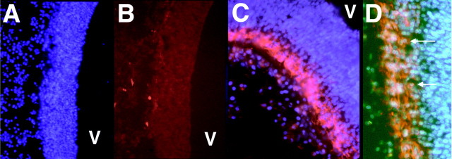
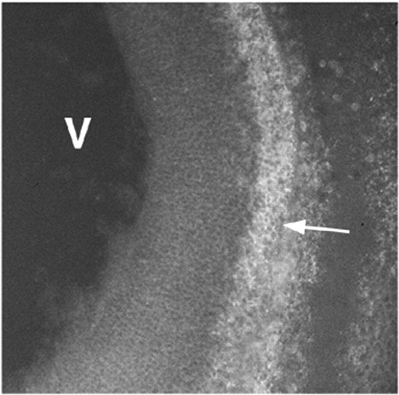
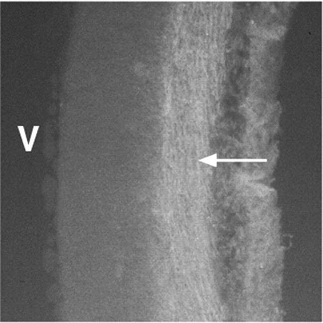
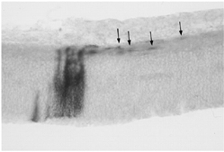
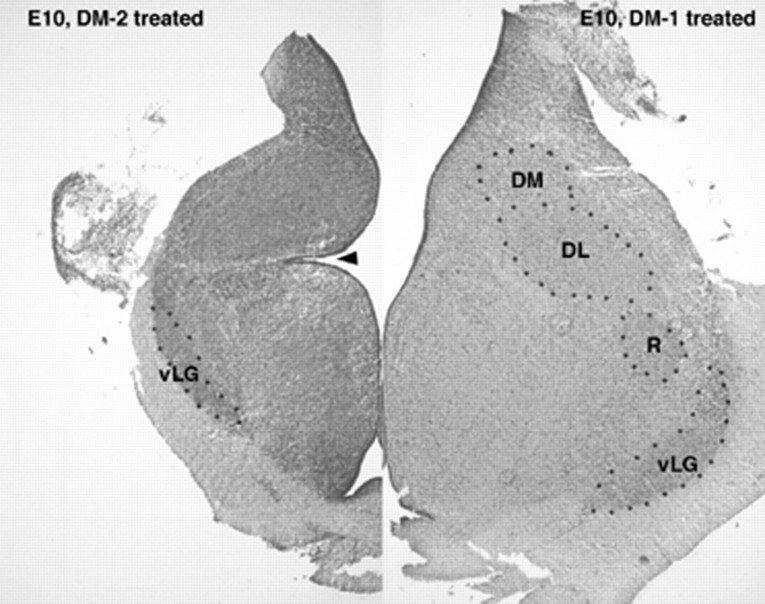
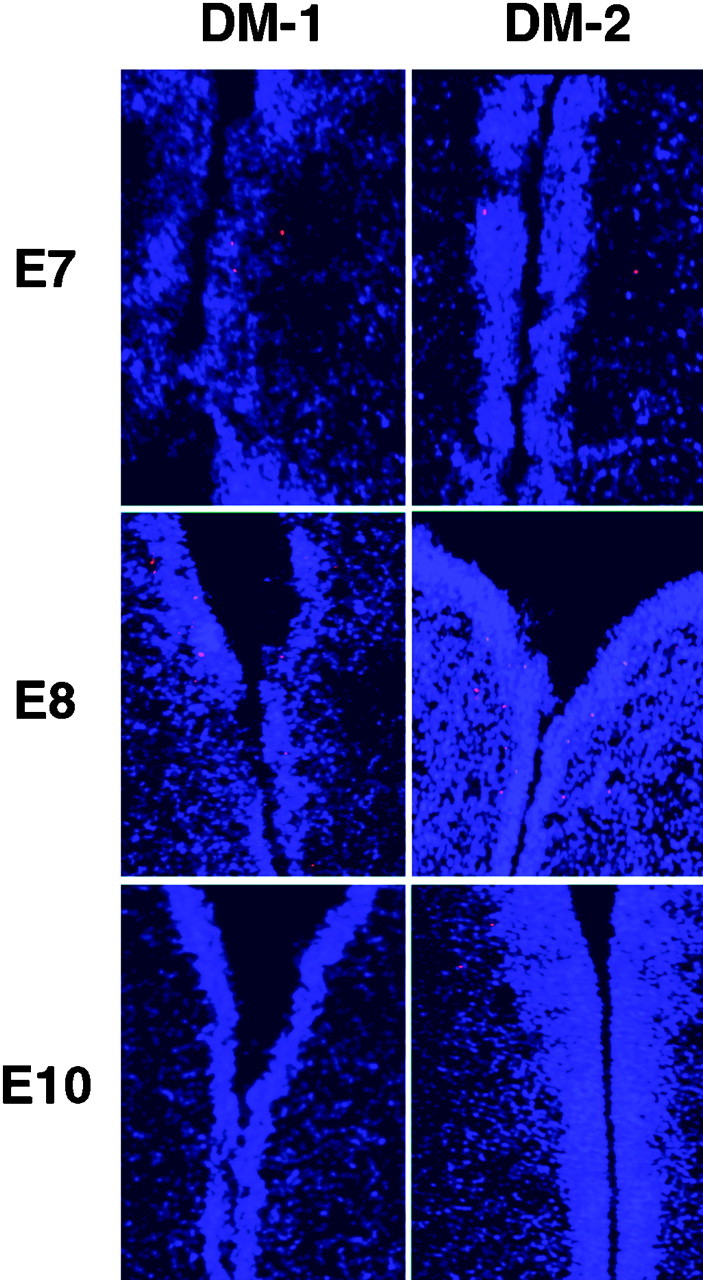
Cell migration is fundamental to normal CNS development. Radial migration, along radial glial fibers, has been the principal pathway studied, however, nonradial or tangential cell migration has increasingly been identified at all levels of the CNS. Receptors, cell adhesion molecules, and extracellular matrix molecules have all been shown to participate in radial cell migration. In contrast, the molecular basis of nonradial cell migration has only recently begun to be elucidated. Using replication defective retroviral vectors we have determined the location and time when nonradial cell migration begins in the developing chick diencephalon. We have identified three molecules that are expressed in spatially and temporally restricted domains that are consistent with them playing a role in nonradial cell migration. One of these molecules, DM-GRASP, a transmembrane protein with five extracellular Ig domains, is expressed on the nonradially migrating cells in addition to axons. To test the hypothesis that DM-GRASP participates in guiding nonradial cell migration, we injected a replication-defective retroviral vector used for lineage tracing followed by a DM-GRASP blocking antibody. Embryos injected with the blocking antibody showed a near complete block in nonradial cell migration specifically where DM-GRASP is expressed. Furthermore, morphological analyses revealed disruption of the normal architecture of the diencephalon indicating nonradial cell migration is necessary for normal morphological development of the brain. Our data indicate that DM-GRASP is necessary for nonradial cell migration in the chick diencephalon and have provided a system to further explore the function of nonradial cell migration during CNS development.


Figures

Fig. 1.
Fig. 1.
Immunofluorescence with an antibody to DM-GRASP/BEN. On E4, no staining is seen in the wall of the diencephalon (A, DAPI staining to show location of diencephalic wall; B, secondary antibody conjugated to Texas Red), although occasionally small axons were seen coursing dorsal to ventral. By E5 DM-GRASP/BEN is expressed at high levels outside the VZ (C, red fluorescence). At higher magnification (D) individual cells are decorated with the DM-1 antibody (arrows). V,Ventricle.
Fig. 2.
Fig. 2.
An embryo intravascularly injected at E6 with DM-2 and harvested 12 hr later shows localization of DM-GRASP/BEN on cells (arrow) outside the VZ (compare with Fig.1c). Sections were treated with secondary antibody conjugated to FITC after fixation and cryosectioning. V,Ventricle.
Fig. 3.
Fig. 3.
Axons were immunolabeled with Tuj1, indicating that DM-2 treatment did not disrupt early axon growth through the diencephalon (arrow points to labeled axons). Embryos were treated on E4 and E5 with DM-2 and evaluated on E7.V, Ventricle.
Fig. 4.
Fig. 4.
Embryos injected with DM-1 or no antibody (A, B) showed clones (identified with the LZ12 retrovirus) with both radial and nonradial cell dispersion. The ventricle is at the bottom (defined by thebottom series of black dots), and the pial surface is at the top (defined by thetop series of black dots) in each image. Radial clones span from the VZ at the bottom of each image toward the top. Nonradial cell dispersion (arrows) was identified in approximately one-third of all clones (A, B). Only radial clones were found in embryos treated with DM-2 (C, D; with two exceptions, see Results).
Fig. 5.
Fig. 5.
Nonradial dispersion of cells (arrows) is observed in the tectum of DM-2-treated embryos where DM-GRASP is not expressed at this developmental time (clones identified by alkaline phosphatase histochemistry after CHAPOL retroviral infection).
Fig. 6.
Fig. 6.
Histological sections of the diencephalon of E10 embryos treated with DM-1 (right) or DM-2 (left). Both sections are taken at approximately the same anatomic level. Embryos injected with DM-1 showed normal diencephalic morphology, whereas those injected with DM-2 had a small diencephalon, loss of neuropil, and poorly organized nuclear groups. This is particularly true for the dorsal nuclei. The ventral nuclei (e.g., ventral lateral geniculate body; vLG) were somewhat better preserved although small, whereas the dorsal nuclei (e.g., nucleus rotundum, R; dorsal lateral,DL; and dorsal medial, DM) nuclei could not be delineated. Furthermore, the sulcus limitans (arrowhead) was extremely deep in DM-2-treated embryos compared to controls or embryos treated with DM-1.
Fig. 7.
Fig. 7.
TUNEL assay on sections through the diencephalon of E7, E8, and E10 embryos after injection of either DM-1 or DM-2. No difference was observed in the pattern or number of TUNEL+ cells between these two experimental groups or those injected with PBS alone. A slight increase in the number of TUNEL-positive cells was observed at E8 in all of the experimental groups. These data likely reflect the normal programmed cell death that occurs in the diencephalon during development.

Similar articles

Cited by

References

    1. Anderson SA, Eisenstat DD, Shi L, Rubenstein JL. Interneuron migration from basal forebrain to neocortex: dependence on Dlx genes. Science. 1997;278:474–476. - PubMed
    1. Anton E, Cameron R, Rakic P. Role of neuron-glial junctional domain proteins in the maintenance and termination of neuronal migration across the embryonic cerebral wall. J Neurosci. 1996;16:2283–2293. - PMC - PubMed
    1. Anton ES, Marchionni MA, Lee KF, Rakic P. Role of GGF/neuregulin signaling in interactions between migrating neurons and radial glia in the developing cerebral cortex. Development. 1997;124:3501–3510. - PubMed
    1. Asou H, Miura M, Kobayashi M, Uyemura K, Itoh K. Cell adhesion molecule L1 guides cell migration in primary reaggregation cultures of mouse cerebellar cells. Neurosci Lett. 1992;144:221–224. - PubMed
    1. Austin CP, Cepko CL. Cellular migration patterns in the developing mouse cerebral cortex. Development. 1990;110:713–732. - PubMed

Publication types

MeSH terms

Substances

LinkOut - more resources