. 2000 Mar 15;20(6):2295-306.
doi: 10.1523/JNEUROSCI.20-06-02295.2000.

The flathead mutation causes CNS-specific developmental abnormalities and apoptosis

Affiliations

The flathead mutation causes CNS-specific developmental abnormalities and apoptosis

M R Roberts et al. J Neurosci. .

Abstract

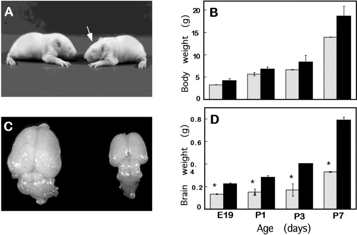
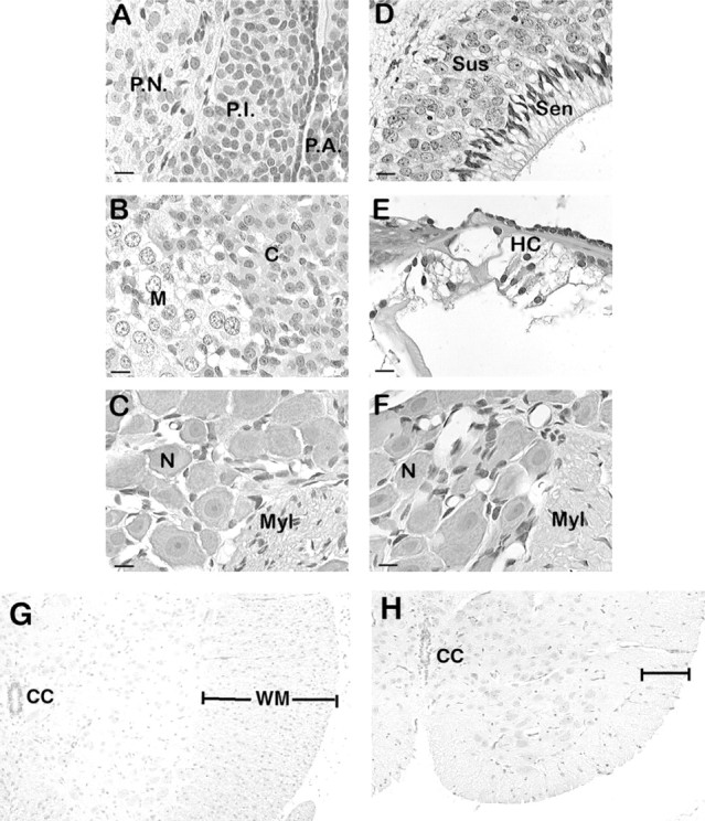
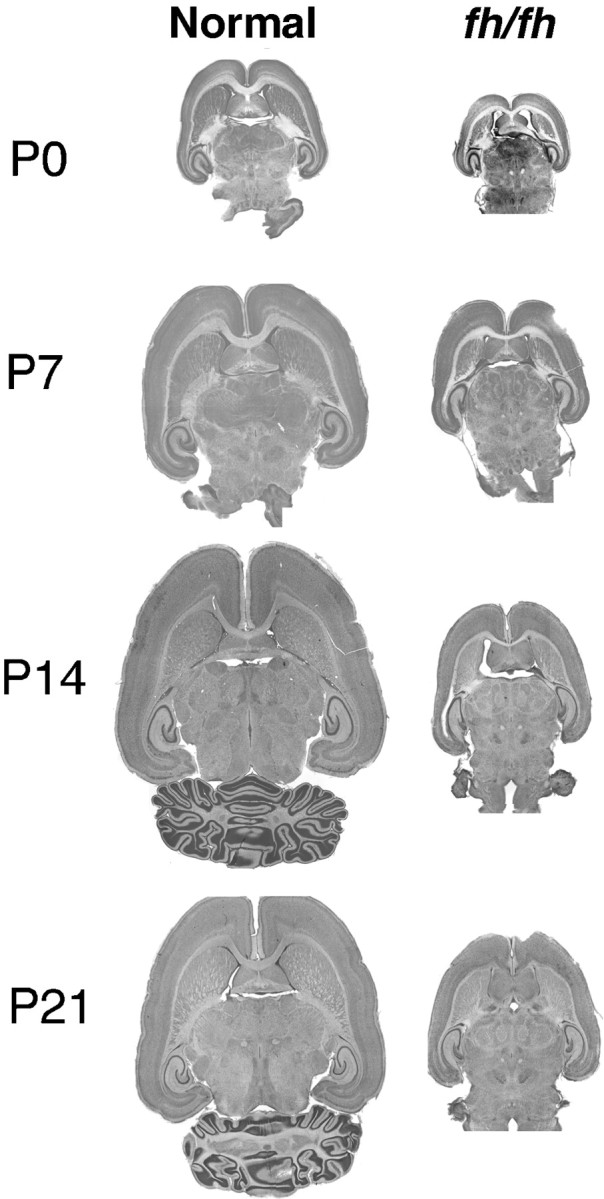
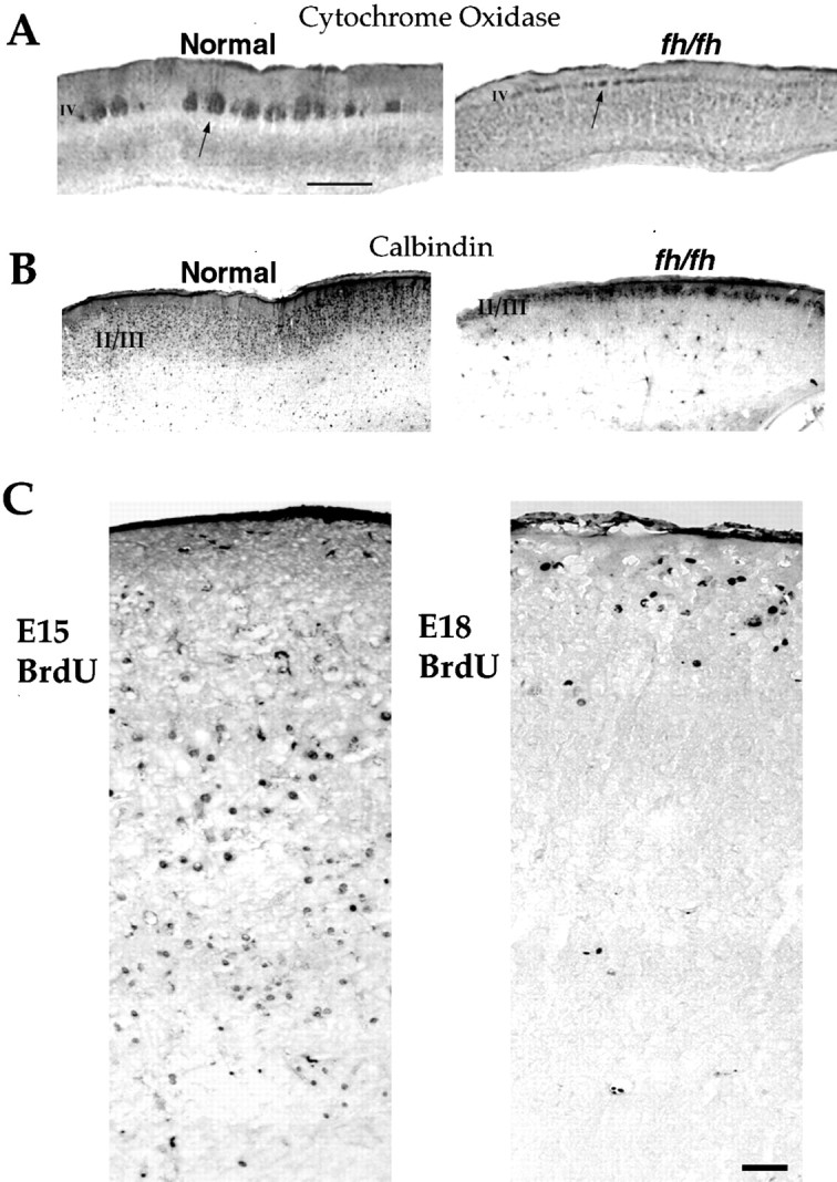
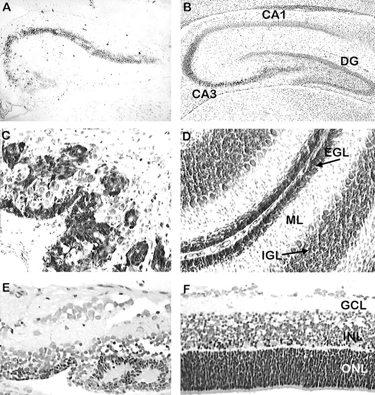
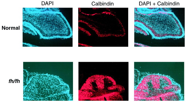
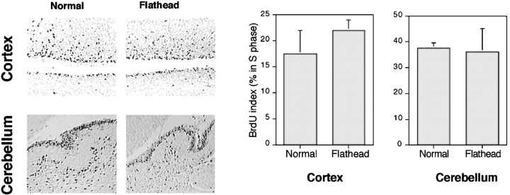
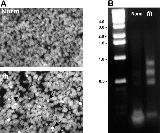
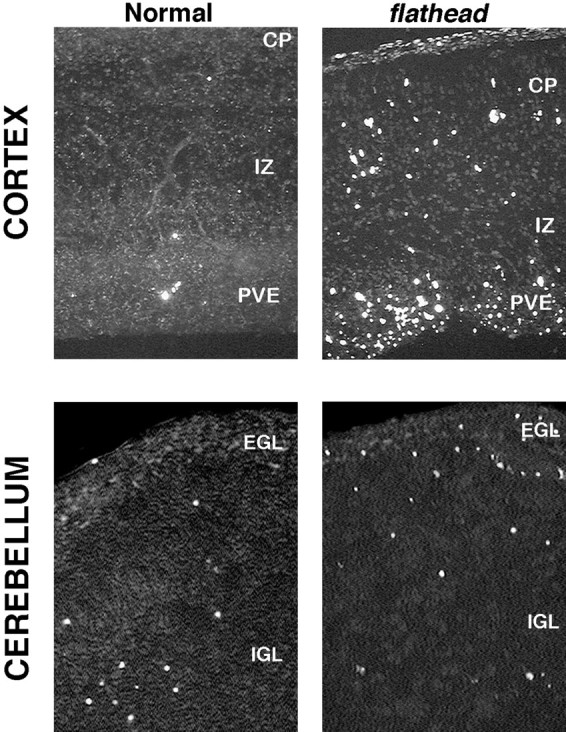
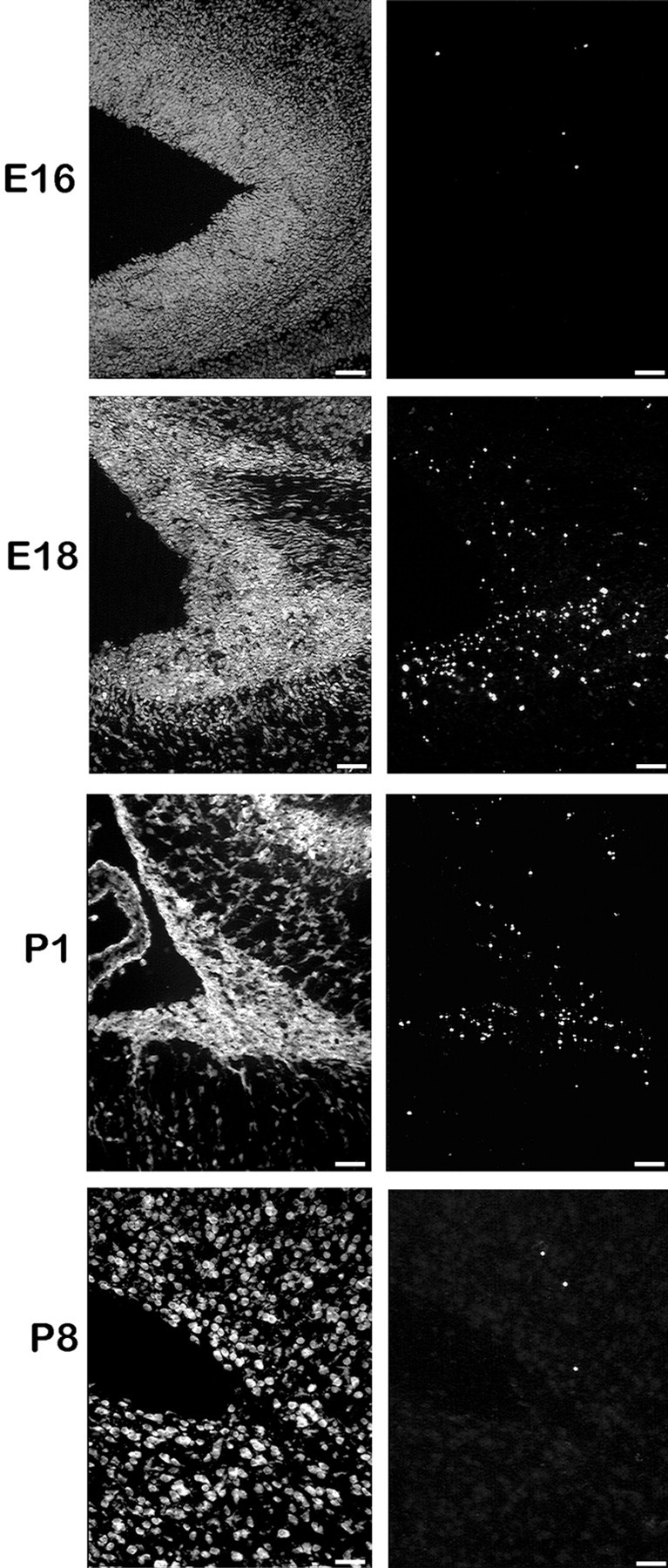
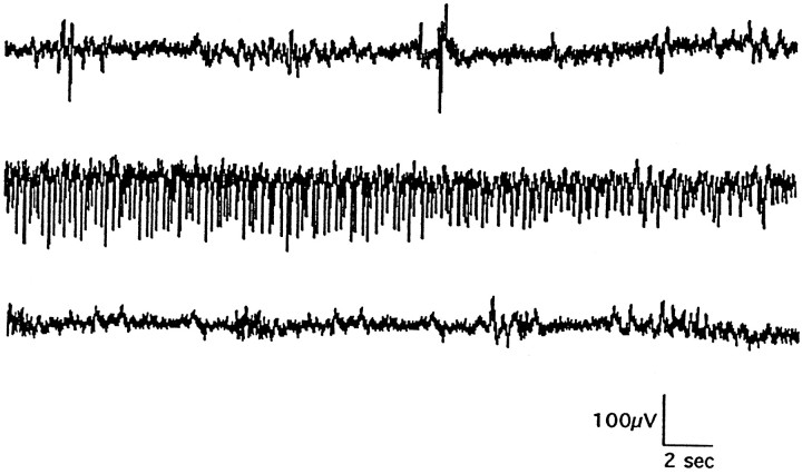
We describe a new mutation, flathead (fh), that arose spontaneously in an inbred colony of Wistar rats. The mutation is autosomal recessive, and the behavioral phenotype of fh/fh rats includes spontaneous seizures, tremor, impaired coordination, and premature death. A striking feature of the fh mutation is a dramatic reduction in brain size (40% of normal at birth). In contrast, no abnormalities are evident in the peripheral nervous system or in other tissues outside of the CNS. Although bromodeoxyuridine incorporation assays indicate that the rate of cell proliferation in the fh/fh cortex is similar to that of unaffected animals, in situ terminal deoxynucleotidyl transferase-mediated dUTP-biotin end-labeling assays reveal a dramatic increase in apoptotic cell death beginning after embryonic day 16 (E16). At E18 there is a 20-fold increase in cell death in the ventricular zone of fh/fh neocortex, and at postnatal day 1 (P1), the number of apoptotic cells is still two times that of normal. However, by P8 the extent of cell death in fh/fh is comparable to that of unaffected littermates, indicating that the reduction in brain growth is caused by abnormally high apoptosis during a discrete developmental period. Late-developing structures such as the cerebellum, neocortex, hippocampus, and retina are most severely affected by the fh mutation. Within these structures, later-generated neuronal populations are selectively depleted. Together, these results suggest that the flathead gene is essential for a developmental event required for the generation and maturation of late-born cell populations in the brain.


Figures

Fig. 1.
Fig. 1.
The flathead mutation preferentially affects the brain. A, The bodies of unaffected (left) and flathead(right) littermates on P10 are similar in size. Also note the slightly flattened curvature of the flatheadskull (arrow). B, A graph of body weights of flathead (striped bars) and unaffected rats (black bars) reveals no significant difference in body weights from E19 until the end of the first postnatal week. C, A flathead brain (right) is significantly smaller than an unaffected (left) littermate on P14. Note that although the entire brain is smaller, the cortex and cerebellum are especially reduced in size. D, A graph of brain weights from E19 to P7 shows that a flathead brain weighs approximately one-half that of a normal brain. Four to eight animals were used for each point. *p < 0.05.
Fig. 2.
Fig. 2.
Light microscopic comparison of body tissues from mutant and unaffected animals did not reveal any differences.fh/fh and unaffected littermates of ages 1, 7, 14, and 21 d were perfused, fixed, and sectioned as described in Materials and Methods. Sections were stained with hematoxylin and eosin and examined by light microscopy. Images from representativefh/fh tissues are shown. A, Pituitary at P14. B, Adrenal gland at P14. C, Dorsal root ganglion at P21. D, Olfactory epithelium at P21.E, Cochlea at P14. F, Superior cervical ganglion at P21. G and H show thoracic spinal cord from normal and fh/fh animals at P14.P.N., Pars nervosa; P.I., pars intermedia; P.A., pars anterior; C, adrenal cortex; M, adrenal medulla; N, neuron; Myl, myelinated nerve fibers;Sus, suscentacular cells; Sen, sensory cells; HC, hair cells; CN, central canal;WM, white matter.
Fig. 3.
Fig. 3.
Light microscopic comparison of horizontal brain sections of fh/fh and unaffected littermates at P0, P7, P14, and P21. Halothane-anesthetized animals were intracardially perfused and then fixed and sectioned on a vibratome at 50 μm. Horizontal sections were mounted on gel-coated slides and stained with cresyl violet. The fh/fh brain is significantly smaller at all ages. Although all brain regions are reduced in size, the cerebral cortex and cerebellum are particularly affected infh/fh.
Fig. 4.
Fig. 4.
fh/fh mutants have reduced thickness of superficial layers but display a normal inside/out pattern of migration in neocortex. A, Cytochrome oxidase stain of somatosensory cortex. Note the thinning and transposition of layer IV toward the pial surface. Scale bar, 1 mm. B, Calbindin immunostaining in fh/fh labels a thin upper layer, which is wider in wild type. C, BrdU immunocytochemistry of a P14 fh/fh animal that was pulsed with BrdU at E15 (left) and another P14fh/fh that was pulsed with BrdU at E18 (right). Labeling was examined in sections 2 weeks after birth. Most of the cells labeled by an E18 injection are localized to upper layers of somatosensory cortex, whereas the E15 injection resulted in staining throughout lower layers of neocortex. Scale bar, 50 μm.
Fig. 5.
Fig. 5.
Thionin staining reveals depletion of neuronal populations born late in neurogenesis and disruption of normal laminar structure. Paraffin-embedded sections (10 μm) of fh/fh (A) and unaffected (B) hippocampus at P21 were stained with thionin and analyzed by light microscopy. The figure shows that the dentate gyrus (DG) is virtually absent in the mutant, and the CA3 region of Ammon's horn is shortened and cell sparse. Sagittal sections of P14 fh/fh(C) and unaffected (D) cerebellum show that some external granule cell layer (EGL) and scattered PCs are present in thefh/fh, but the internal granule cell layer (IGL) is absent. Sections of the P12 retina fromfh/fh (E) and unaffected (F) show that the photoreceptors in the outer nuclear layer (ONL) are severely depleted. Although the laminar structure is disrupted, the inner nuclear layer (INL) and ganglion cell layer (GCL) are less affected.
Fig. 6.
Fig. 6.
Calbindin immunostaining reveals thatfh/fh cerebellum contains mostly Purkinje neurons. Frozen sagittal sections of 7-d-old normal and fh/fhbrains were processed for immunohistological detection of calbindin, a protein expressed specifically by Purkinje neurons in the cerebellum.Red represents calbindin staining, whereasblue represents nuclei stained with DAPI.
Fig. 7.
Fig. 7.
BrdU pulse labeling reveals a comparable rate of proliferation in fh/fh and unaffected cortex. Animals were injected with 60 μg/g of BrdU 1 hr before they were killed. Paraffin sections were processed for immunohistological detection of BrdU incorporation as described in Materials and Methods. BrdU labeling in the E19 unaffected (left) and fh/fh(right) cortex and P1 cerebellum is shown. The percentage of total BrdU-positive cells in the PVE of the cerebral cortex and EGL of the cerebellum has been quantified (n = 4) and shown graphically. No significant difference in the proportion of BrdU-positive cells was observed in cortical or cerebellar sections between unaffected andfh/fh animals.
Fig. 8.
Fig. 8.
Increased cell death in fh/fhcortex. Cell death was examined by nuclear staining (A) and LM-PCR amplification of DNA (B). DAPI staining reveals a greater number of pyknotic nuclei in the fh/fh cortex (arrows) than in the unaffected cortex at E18. To determine whether these pyknotic nuclei might be apoptotic (rather than necrotic or mitotic), we used the LM-PCR technique to preferentially amplify fragmented DNA from E18 fh/fh and normal brains. After amplification, DNA from unaffected and fh/fhanimals was analyzed by electrophoresis, which revealed a laddering pattern in DNA from the fh/fh but not the unaffected brain. Numbers on the left indicate molecular sizes in kilobases.
Fig. 9.
Fig. 9.
TUNEL-labeling reveals an increase of apoptotic cell death in the fh/fh cortex and cerebellum. Frozen tissue was sectioned at 10 μm, permeabilized with proteinase K, and incubated with florescein-conjugated terminal transferase for 1 hr to label fragmented DNA. Top panels, TUNEL labeling in the E18 unaffected (left) and fh/fh(right) cortex. Although the number of dying cells is dramatically increased in all areas of the cortex at E18, the pattern of cell death in the fh/fh cortex mirrors the pattern observed in the normal cortex at this age. Specifically, the areas of highest cell death occur in the periventricular epithelium (PVE), which contains mostly proliferating cells, whereas the cortical plate (CP) and intermediate zone (IZ) have less cell death. Bottom panels, TUNEL labeling in the unaffected (left) andfh/fh (right) cerebellum at P1. The external granule layer (EGL) of fh/fhdisplays substantially more TUNEL-positive cells than that of an unaffected littermate. Although a distinct internal granule layer (IGL) is lacking in the fh/fh cerebellar, the proportion of TUNEL-positive cells in the region internal to the EGL was comparable to that of the normal IGL.
Fig. 10.
Fig. 10.
TUNEL labeling of the ventricular region of the cerebral cortex at E16, E18, P1, and P8 reveals that cell death occurs within a narrow time window during development of thefh/fh cortex. Frozen tissue was sectioned at 10 μm, rinsed in xylenes to remove lipids, permeabilized with proteinase K, and incubated with florescein-conjugated terminal transferase to label fragmented DNA. Sections were counterstained with propidium iodide (seepanels at left). Our results indicate that the onset and peak of cell death occurs around E18. Although thefh/fh cortex has a higher rate of cell death than a normal brain at P1, the frequency of cell death is approximately one-half of what it was at E18. By P8, only a few scattered cells are TUNEL positive in the fh/fh, which is similar to the pattern observed in a normal brain (data not shown).
Fig. 11.
Fig. 11.
Representative EEG recording from afh/fh rat at P14 before (top), during (middle), and after (bottom) a spontaneous convulsive seizure. Such seizure activity has been recorded in 72 animals, with seizures occurring at a rate of four to six per hour from P7 to P18.

Similar articles

Cited by

References

    1. Alder J, Cho NK, Hatten ME. Embryonic precursor cells from the rhombic lip are specified to a cerebellar granule neuron identity. Neuron. 1996;17:389–399. - PubMed
    1. Altman J, Bayer SA. Embryonic development of the rat cerebellum. I. Delineation of the cerebellar primordium and early cell movements. J Comp Neurol. 1985a;231:1–26. - PubMed
    1. Altman J, Bayer SA. Embryonic development of the rat cerebellum. III. Regional differences in the time of origin, migration, and settling of Purkinje cells. J Comp Neurol. 1985b;231:42–65. - PubMed
    1. Angevine JB, Sidman RL. Autoradiographic study for cell migration during histogenesis of the cerebral cortex in the mouse. Nature. 1961;192:766–768. - PubMed
    1. Bayer SA. Development of the hippocampal region in the rat. II. Morphogenesis during embryonic and early postnatal life. J Comp Neurol. 1980;190:115–134. - PubMed

MeSH terms

LinkOut - more resources