

Concurrent MRI and diffuse optical tomography of breast after indocyanine green enhancement
- PMID: 10706610
- PMCID: PMC16004
- DOI: 10.1073/pnas.040570597
Concurrent MRI and diffuse optical tomography of breast after indocyanine green enhancement
Abstract
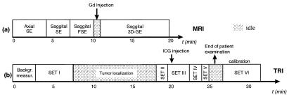
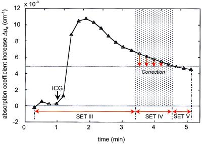
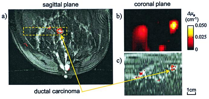
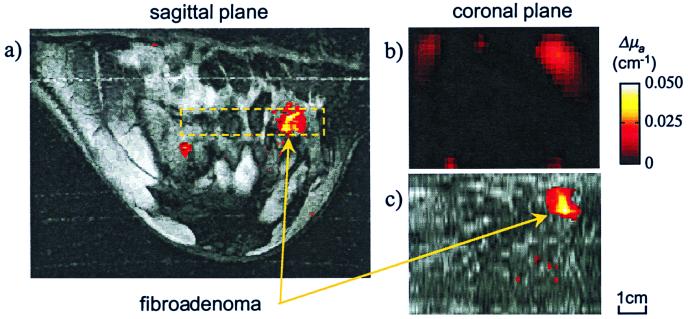
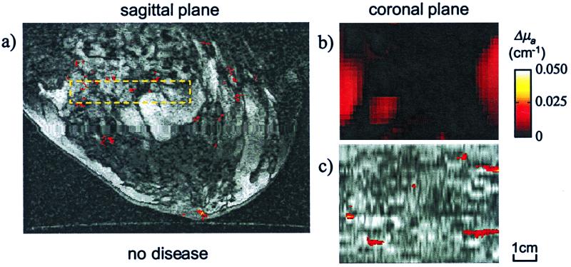
We present quantitative optical images of human breast in vivo. The images were obtained by using near-infrared diffuse optical tomography (DOT) after the administration of indocyanine green (ICG) for contrast enhancement. The optical examination was performed concurrently with a magnetic resonance imaging (MRI) exam on patients scheduled for excisional biopsy or surgery so that accurate image coregistration and histopathological information of the suspicious lesions was available. The ICG-enhanced optical images coregistered accurately with Gadolinium-enhanced magnetic resonance images validating the ability of DOT to image breast tissue. In contrast to simple transillumination, we found that DOT provides for localization and quantification of exogenous tissue chromophore concentrations. Additionally our use of ICG, an albumin bound absorbing dye in plasma, demonstrates the potential to differentiate disease based on the quantified enhancement of suspicious lesions.
Figures
References
-
- Arridge S R. Appl Opt. 1995;34:7395–7409. - PubMed
-
- O'Leary M A, Boas D A, Chance B, Yodh A G. Opt Lett. 1995;20:426–428. - PubMed
-
- Jiang H, Paulsen K D, Osterberg U L, Pogue B W, Patterson M S. J Opt Soc Am A. 1996;13:253–266.
-
- Gonatas C P, Ishii M, Leigh J S, Schotland J C. Phys Rev E. 1995;52:4361–4365. - PubMed
-
- Chang J H, Graber H L, Barbour R L. IEEE T Bio Med Eng. 1997;44:810–822. - PubMed
Publication types
MeSH terms
Substances
Grants and funding
LinkOut - more resources
Full Text Sources
Other Literature Sources
Medical
