. 2000 Mar 14;97(6):2609-14.
doi: 10.1073/pnas.040576497.

Notch signaling directly controls cell proliferation in the Drosophila wing disc

Affiliations

Notch signaling directly controls cell proliferation in the Drosophila wing disc

A Baonza et al. Proc Natl Acad Sci U S A. .

Abstract

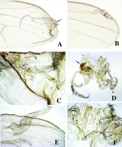
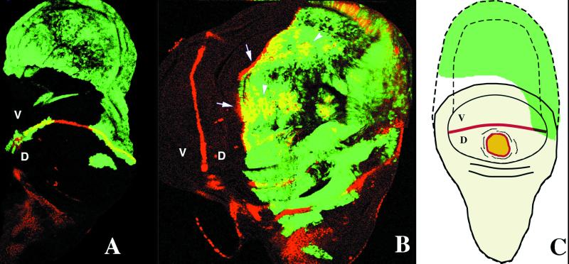
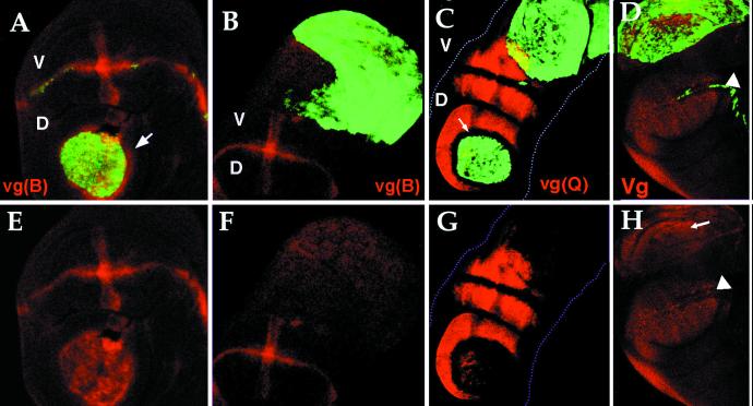
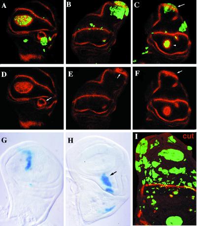
Notch signaling is involved in cell differentiation and patterning during morphogenesis. In the Drosophila wing, Notch activity regulates the expression of several genes at the dorsal/ventral boundary, and this is thought to elicit wing-cell proliferation. In this work, we show the effect of clones of cells expressing different forms of several members of the Notch signaling pathway, which result in an alteration of Notch activity. The ectopic expression in clones of activated forms of Notch or of its ligands (Delta or Serrate) in the wing causes outgrowths associated with the appearance of ectopic wing margins. These outgrowths consist of mutant territories and of surrounding wild-type cells. However, the ectopic expression of Delta, at low levels in ventral clones, causes large outgrowths that are associated neither with the generation of wing margin structures nor with the expression of genes characteristic of the dorsal/ventral boundary. These results suggest that Notch activity is directly involved in cell proliferation, independently of its role in the formation of the dorsal/ventral boundary. We propose that the nonautonomous effects (induction of extraproliferation and vein differentiation in the surrounding wild-type cells) result from pattern accommodation to positional values caused by the ectopic expression of Notch.


Figures

Figure 1
Figure 1
Adult phenotypes caused by clones of ectopic expression of Nintra using the abx/Ubx promoter (A and B) and Dl by using the actin promoter (C–F). In A and B, clones of Nintra-expressing cells (forked) in the wing blade cause the ectopic differentiation of wing margin structures (arrows) and induce the proliferation of wild-type cells surrounding the clone (dotted line). The clones generated close to the a/p boundary and away from the d/v boundary cause larger outgrowths (A) than clones close to the d/v border (B). In C, a clone (red dotted line) of Dl-expressing cells (yellow) in the dorsal compartment causes wing outgrowths that contain wing margin structures at the clone boundaries (arrows indicate mutant elements). (D) Large outgrowths in the legs associated with clones of Dl-expressing cells (arrow indicates a large outgrowth in the coxal region and arrowhead, an enlarged leg). In E, clones of Dl-expressing cell in the ventral compartment of the wing cause large outgrowth that is not associated with the differentiation of wing margin structures. The wild-type cells constitute only a small fraction of the outgrowth (dotted line). (F) Large outgrowth (dotted line) in the notum caused by a clone (arrows) of Dl-expressing cells.
Figure 2
Figure 2
Effects of clones of Dl-expressing cells (green, GFP labeled) on the expression of CT (antibody against CT, staining in red) in third instar imaginal wing discs in the dorsal (D) and ventral (V) compartments. In A, ventral clones cause large outgrowths where CT is not ectopically expressed. In B, dorsal clones of Dl-expressing cells induce the ectopic expression of CT in some cells within the clone (arrowheads) and in the wild-type cells surrounding the clone (arrows). In C, schematic representation of the effects caused by clones of Dl-expressing cells. Ventral clones that do not express CT cause large outgrowth, and dorsal clones cause the ectopic expression of CT within the clone (yellow) and in adjacent wild-type cells (red).
Figure 3
Figure 3
Effect of clones of Dl-expressing cells (green, GFP) on the expression of vestigial boundary enhancer [vg(B)] red in (A, B, E, and F), quadrant enhancer [vg(Q)] red in (C and G), and pattern of VG protein monitored by using an antibody against VG (D and H). In A and E, the vestigial boundary enhancer [vg(B)] is activated in the outgrowth caused by a dorsal clone in both mutant and wild-type cells surrounding the clone (arrow), but not in ventral clones (B and F). In C and G, the Quadrant Enhancer [vg(Q)] is activated in the outgrowth caused by Dl dorsal clones but not in ventral. In dorsal clones the vg(Q) enhancer is repressed in wild-type cells adjacent to the clone but ectopically activated in both mutant cells and wild-type cells [arrow indicates the wild-type cells adjacent to the clone that not express vg(Q) enhancer]. In D and H, the entire wing pouch, including the outgrowth, expresses the VG protein (arrow) (arrowheads point to the d/v boundary). The cells in the outgrowth that do not show VG expression are in a different focal plane.
Figure 4
Figure 4
Expression pattern of WG (A–F) and CT (I) (red) in wing discs with clones of Dl-expressing cells (green, GFP); clones of wg-expressing cells (G and H) (blue, β-galactosidase). In A–F, different examples of clones of Dl-expressing cells. In A, C, D, and F, dorsal clones express high levels of WG within the clone as well as in the wild-type cells adjacent to the clone (arrowhead in C). In B, C, E, and F, the clones in the ventral compartment that abut the external and internal rings of WG expression cause large outgrowths with elongation of the internal ring of WG expression (arrows in C). High levels of WG in some cells within the clone can be observed (arrows in E and F). In G and H, clones of wg-expressing cells in the wing blade never cause outgrowths, whereas they do in notum and wing base (H). In I, compare the small size of clones of Dl-expressing cells closer to the d/v boundary with the large ones in the proximal region of the wing.

Similar articles

Cited by

References

    1. Artavanis-Tsakonas S, Rand M D, Lake R J. Science. 1999;284:770–776. - PubMed
    1. Knust E, Schrons H, Grawe F, Campos-Ortega J A. Genetics. 1992;132:505–518. - PMC - PubMed
    1. Schrons H, Knust E, Campos-Ortega J A. Genetics. 1992;132:481–503. - PMC - PubMed
    1. Lecourtois M, Schweisguth F. Genes Dev. 1995;9:2598–2608. - PubMed
    1. Artavanis-Tsakonas S, Matsuno K, Fortini M E. Science. 1995;268:225–232. - PubMed

Publication types

MeSH terms

LinkOut - more resources