. 2000 Mar 14;97(6):2910-5.
doi: 10.1073/pnas.040577797.

Hyperphosphorylated tau and neurofilament and cytoskeletal disruptions in mice overexpressing human p25, an activator of cdk5

Affiliations

Hyperphosphorylated tau and neurofilament and cytoskeletal disruptions in mice overexpressing human p25, an activator of cdk5

M K Ahlijanian et al. Proc Natl Acad Sci U S A. .

Abstract

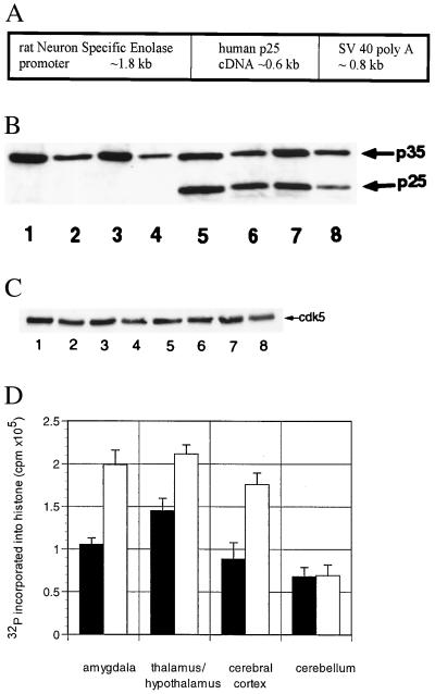
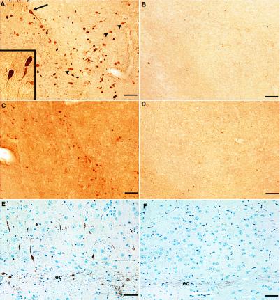
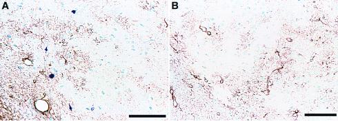
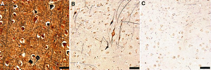
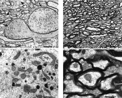
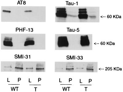
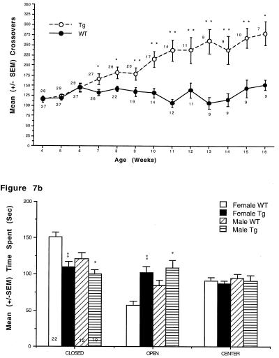
Hyperphosphorylation of microtubule-associated proteins such as tau and neurofilament may underlie the cytoskeletal abnormalities and neuronal death seen in several neurodegenerative diseases including Alzheimer's disease. One potential mechanism of microtubule-associated protein hyperphosphorylation is augmented activity of protein kinases known to associate with microtubules, such as cdk5 or GSK3beta. Here we show that tau and neurofilament are hyperphosphorylated in transgenic mice that overexpress human p25, an activator of cdk5. The p25 transgenic mice display silver-positive neurons using the Bielschowsky stain. Disturbances in neuronal cytoskeletal organization are apparent at the ultrastructural level. These changes are localized predominantly to the amygdala, thalamus/hypothalamus, and cortex. The p25 transgenic mice display increased spontaneous locomotor activity and differences from control in the elevated plus-maze test. The overexpression of an activator of cdk5 in transgenic mice results in increased cdk5 activity that is sufficient to produce hyperphosphorylation of tau and neurofilament as well as cytoskeletal disruptions reminiscent of Alzheimer's disease and other neurodegenerative diseases.


Figures

Figure 1
Figure 1
(A) NSE-p25 transgene. Expression construct for human p25 cDNA sequences under the control of the rat NSE promoter. (B) Expression of p25 in transgenic mice. Western blots of wild-type (lanes 1–4) and transgenic (lanes 5–8) brain lysates from the amygdala (lanes 1 and 5), thalamus/hypothalamus (lanes 2 and 6), cerebral cortex (lanes 3 and 7), and cerebellum (lanes 4 and 8) using an antibody that recognizes both p35 and p25. (C) Expression of cdk5 in transgenic mice. Immunoblots of wild-type (lanes 1–4) and transgenic (lanes 5–8) brain lysates from the amygdala (lanes 1 and 5), thalamus/hypothalamus (lanes 2 and 6), cerebral cortex (lanes 3 and 7), and cerebellum (lanes 4 and 8) using an antibody that recognizes cdk5. (D) Increased cdk5 activity in transgenic mice. cdk5/p25/p35 complexes from wild-type (■) and transgenic (○) amygdala, thalamus/hypothalamus, cerebral cortex, and cerebellum were immunoprecipitated with an antibody that recognizes cdk5, then assessed for kinase activity by incubating with [32P]ATP and histone. The y axis represents the total cpm incorporated into histone minus a blank (no primary antibody) for three mice in each group (mean ± SEM). Similar results were obtained for an antibody that recognizes p35/p25 (not shown).
Figure 2
Figure 2
Abnormal phosphorylation epitopes in p25 transgenic vs. wild-type brains. Brain sections from p25 transgenic animals (A, C, and E) were immunostained in parallel with corresponding regions from wild-type animals (B, D, and F). Immunostaining of AT8 in transgenic (A) and wild-type (B) amygdala. Many cells in this region had densely stained cell bodies with well defined immunopositive axons and axonal hillocks (arrow). Inset shows an increased magnification of these cells from a different transgenic animal. Other immunopositive structures were more lightly stained and had diffuse borders (arrowheads). Immunostaining of PHF13 in transgenic (C) and wild-type (D) amygdala is shown. Background staining of this antibody was routinely higher in transgenic brain sections and represents greater immunoreactivity in the neuropil. Immunostaining with SMI34 in cortex adjacent to external capsule in transgenic (E) and wild-type (F) mice also is shown. Note the constitutive staining of external capsule axons (ec) by SMI34 in both transgenic and wild-type sections. This pattern of staining was seen in 14 of 15 4-month-old transgenic mice and never seen in 6 wild-type mice of the same age. (Bar = 50 μ.)
Figure 3
Figure 3
Comparison of AT8 and GFAP immunostaining. Amygdala sections from transgenic (A) and wild-type mice (B) were immunostained with AT8 (blue) and an anti-GFAP antibody (brown). Note the nonoverlapping distribution of GFAP and AT8 staining in the transgenic section (A) and the lack of AT8 staining in the wild-type section (B). (Bar = 100 μ.)
Figure 4
Figure 4
Argyrophilic structures from p25 transgenic brain. A section from cortex of an Alzheimer's brain (A) is compared with sections from p25 transgenic cortex (B) and wild-type cortex (C) silver-stained by using the modified Bielschowsky method. Unlike wild type, p25 transgenic brain contained argyrophilic cell bodies and axons. This pattern of staining was seen in 14 of 15 4-month-old transgenic and never seen in 15 wild-type mice of the same age. (Bar = 50 μ.)
Figure 5
Figure 5
Electron microscopy of amygdala from p25 transgenic and wild-type brains. Axons within the amygdala of transgenic mice were markedly dilated (A). Compare abnormal dilated axons (arrows) with axons of normal size (arrowheads). Mitochondria (M) and lysosomes (L) were clustered together abnormally and were interspersed among disorganized cytoskeletal components (B, *). Axons from the same location in age-matched, wild-type mice were normal (C and D). Axonal changes were observed ultrastructurally in five of six transgenic mice aged 3–6 months and not observed in any of five age-matched, wild-type mice. [Bars = 4 μ (A and C) or 500 nm (B and D).]
Figure 6
Figure 6
Immunoblots for tau and neurofilament. Immunoblots containing tissue lysates (L) and pellets (P) derived from the amygdala from wild-type (WT) and p25 transgenic (P) mice were probed with antibodies specific for phospho- (AT8) and dephospho-Thr-202, Ser-205 of tau (Tau-1), phospho-tau Ser-396 (PHF-13), total tau (Tau-5), phospho-neurofilament (SMI-31), and total neurofilament (SMI-33). No differences in any of these epitopes between transgenic and wild-type mice were detected.
Figure 7
Figure 7
(A) p25 transgenic mice exhibit increased locomotor activity in an open field. p25 transgenic mice exhibited significantly more square crossings than wild-type littermates in an open field (*, P < .05; **, P < .01). Females (data not shown) showed the same effect beginning at 5 weeks of age. Numbers next to symbols represent the number of animals tested at each time point. (B) p25 transgenic mice spend increased time on the open arms of an elevated plus maze. p25 transgenic mice exhibited significantly greater time than wild-type littermates on the open arms of an elevated plus maze (*, P < .05; **, P < .01). Numbers within bars represent the number of animals in each group.

Similar articles

Cited by

References

    1. Goedert M, Crowther R A, Spillantini M G. Neuron. 1998;21:955–958. - PubMed
    1. Imahori K, Uchida T. J Biochem. 1997;121:179–188. - PubMed
    1. Ishiguro K, Takamatsu M, Tomizawa K, Omori A, Takahashi M, Arioka M, Uchida T, Imahori K. J Biol Chem. 1992;267:10897–10901. - PubMed
    1. Hosoi T, Uchiyama M, Okumura E, Saito T, Ishiguro K, Uchida T, Okuyama A, Kishimoto T, Hisanaga S. J Biochem. 1995;117:741–740. - PubMed
    1. Wagner U, Utton M, Gallo J M, Miller C C. J Cell Sci. 1996;109:1537–1543. - PubMed

MeSH terms

LinkOut - more resources