. 2000 Mar 14;97(6):2846-51.
doi: 10.1073/pnas.050569397.

Stress-induced enhancement of skin immune function: A role for gamma interferon

Affiliations

Stress-induced enhancement of skin immune function: A role for gamma interferon

F S Dhabhar et al. Proc Natl Acad Sci U S A. .

Abstract

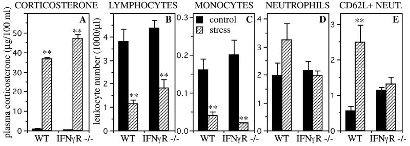
Contrary to the widespread belief that stress is necessarily immunosuppressive, recent studies have shown that, under certain conditions, stress can induce a significant enhancement of a skin cell-mediated immune response [delayed-type hypersensitivity (DTH) or contact hypersensitivity]. Adrenal stress hormones and a stress-induced trafficking of leukocytes from the blood to the skin have been identified as systemic mediators of this immunoenhancement. Because gamma interferon (IFNgamma) is an important cytokine mediator of DTH, the studies described here were designed to examine its role as a local mediator of the stress-induced enhancement of skin DTH. The effect of acute stress on skin DTH was examined in wild-type and IFNgamma receptor-deficient (IFNgammaR-/-) mice that had previously been sensitized with 2,4-dinitro-1-fluorobenzene. Acutely stressed wild-type mice showed a significantly larger DTH response than nonstressed mice. In contrast, IFNgammaR-/- mice failed to show a stress-induced enhancement of skin DTH. Immunoneutralization of IFNgamma in wild-type mice significantly reduced the stress-induced enhancement of skin DTH. In addition, an inflammatory response induced by direct IFNgamma administration to the skin was significantly enhanced by acute stress. Our results suggest that IFNgamma is an important local mediator of a stress-induced enhancement of skin DTH. These studies are clinically relevant because, depending on the nature of the antigen, DTH reactions mediate numerous protective (e.g., resistance to viral, bacterial, parasitic, and fungal infections) or pathological (e.g., autoimmune reactions and contact sensitivity reactions such as that to poison ivy) immune responses.


Figures

Figure 1
Figure 1
Mechanisms by which acute stress may enhance skin immunity. Stage 1: The brain detects the presence of a stressor and informs/warns the body by releasing stress hormones. Stage 2: Stress hormones increase the affinity/expression of adhesion molecules on leukocytes and/or endothelial cells. Stage 3: This results in a selective margination of leukocytes within the vasculature of organs such as the skin. Stage 4a: Upon cessation of stress, and in the absence of immunologic challenge at the site of leukocyte margination, leukocytes demarginate and join the circulating leukocyte pool (14). Stage 4b: However, if the stressor is accompanied by an immune challenge at the site of leukocyte margination, leukocytes are recruited by chemokines and cytokines released at the site of antigen entry. Thus, a stressed organism, by virtue of having higher numbers of leukocytes available for recruitment at potential sites of challenge, may mount a more robust immune response than an unstressed organism. Stage 5: In addition to affecting leukocyte distribution, acute stress may increase immunopreparedness by increasing inflammatory mediator production/function and by enhancing antigen presentation, leukocyte recruitment, leukocyte activation, and effector cell function within the site of an immune reaction. Thus, stress hormones and changes in leukocyte distribution within the body may be systemic mediators, and cytokines and chemokines local mediators, of a stress-induced enhancement of immune function.
Figure 2
Figure 2
Effects of stress on the DTH response of wild-type (A) and IFNγR−/− (B) mice. Control animals were left undisturbed while stressed animals were restrained for 2.5 h. The right pinnae of all animals were then challenged with DNFB (0.2%). A time course of changes in the thickness of challenged pinnae is shown. Acute stress significantly enhanced the DTH response of wild-type mice (n = 5, *, P < 0.05; **, P < 0.005, independent t test). In contrast, IFNγR−/− mice failed to show a stress-induced enhancement of skin DTH.
Figure 3
Figure 3
Stress-induced changes in plasma corticosterone levels (A) and blood leukocyte numbers (B–E). Control animals were left undisturbed while stressed animals were restrained for 2.5 h. Wild-type (WT) and IFNγR−/− mice showed a robust stress-induced increase in plasma corticosterone (n = 3, A), and a robust stress-induced decrease in blood lymphocyte (B) and monocyte (C) numbers (n = 5). Wild-type mice also showed a stress-induced increase in blood neutrophil numbers (n = 5, D), which was particularly prominent for CD62L+ neutrophils (E). IFNγR−/− mice failed to show this stress-induced neutrophilia (n = 5). Statistically significant differences are indicated (**, P < 0.005, independent t test).
Figure 4
Figure 4
Effects of immunoneutralization of IFNγ on a stress-induced enhancement of skin DTH in wild-type mice. Animals were injected with rat IgG (A) or anti-IFNγ (B). Then, 1.5 h after antibody administration, control animals were left undisturbed, while stressed animals were restrained for 2.5 h. The right pinnae of all animals were then challenged with DNFB (0.2%). A time course of changes in the thickness of challenged pinnae is shown. Vehicle-injected wild-type mice showed a significant stress-induced enhancement of skin DTH (n = 5; *, P < 0.05, independent t test). In contrast, wild-type mice injected with anti-IFNγ antibody failed to show this enhancement (n = 3).
Figure 5
Figure 5
Effects of acute stress on an inflammatory response to direct administration of IFNγ to the skin of wild-type animals. Animals were not sensitized for this experiment. Control animals were left undisturbed, while stressed animals were restrained for 2.5 h. Mouse recombinant IFNγ was then administered to the right pinnae of all animals. A time course of changes in pinna thickness is shown. Stressed animals showed a significant enhancement of inflammation at the site of IFNγ administration (n = 9; *, P < 0.05). Stressed animals also showed an enhancement of inflammation after saline administration, although the overall response to saline was lower than that observed following IFNγ administration (n = 9; *, P < 0.05).

Similar articles

Cited by

References

    1. Dhabhar F S, McEwen B S. Brain Behav Immun. 1997;11:286–306. - PubMed
    1. Dhabhar F S, McEwen B S. Proc Natl Acad Sci USA. 1999;96:1059–1064. - PMC - PubMed
    1. Dhabhar F S, McEwen B S. J Immunol. 1996;156:2608–2615. - PubMed
    1. McEwen B S. N Engl J Med. 1998;338:171–179. - PubMed
    1. Khansari D N, Murgo A J, Faith R E. Immunol Today. 1990;11:170–175. - PubMed

Publication types

LinkOut - more resources