

Structures of recombinant human and mouse NAD(P)H:quinone oxidoreductases: species comparison and structural changes with substrate binding and release
- PMID: 10706635
- PMCID: PMC16212
- DOI: 10.1073/pnas.97.7.3177
Structures of recombinant human and mouse NAD(P)H:quinone oxidoreductases: species comparison and structural changes with substrate binding and release
Abstract
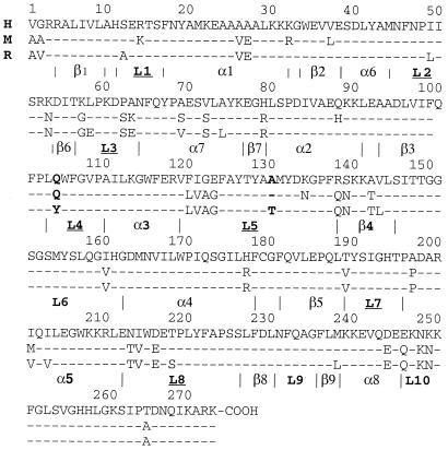
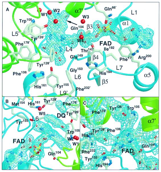
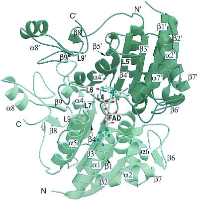
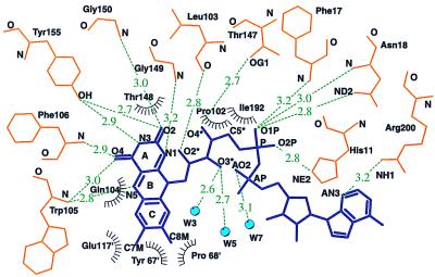
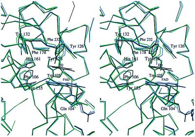
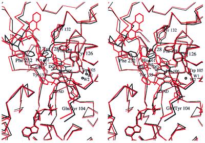
NAD(P)H/quinone acceptor oxidoreductase (QR1, NQO1, formerly DT-diaphorase; EC ) protects animal cells from the deleterious and carcinogenic effects of quinones and other electrophiles. In this paper we report the apoenzyme structures of human (at 1.7-A resolution) and mouse (2.8 A) QR1 and the complex of the human enzyme with the substrate duroquinone (2.5 A) (2,3,5, 6-tetramethyl-p-benzoquinone). In addition to providing a description and rationale of the structural and catalytic differences among several species, these structures reveal the changes that accompany substrate or cofactor (NAD) binding and release. Tyrosine-128 and the loop spanning residues 232-236 close the binding site, partially occupying the space left vacant by the departing molecule (substrate or cofactor). These changes highlight the exquisite control of access to the catalytic site that is required by the ping-pong mechanism in which, after reducing the flavin, NAD(P)(+) leaves the catalytic site and allows substrate to bind at the vacated position. In the human QR1-duroquinone structure one ring carbon is significantly closer to the flavin N5, suggesting a direct hydride transfer to this atom.
Figures
Similar articles
-
Unexpected genetic and structural relationships of a long-forgotten flavoenzyme to NAD(P)H:quinone reductase (DT-diaphorase).Proc Natl Acad Sci U S A. 1997 Mar 4;94(5):1669-74. doi: 10.1073/pnas.94.5.1669. Proc Natl Acad Sci U S A. 1997. PMID: 9050836 Free PMC article.
-
The three-dimensional structure of NAD(P)H:quinone reductase, a flavoprotein involved in cancer chemoprotection and chemotherapy: mechanism of the two-electron reduction.Proc Natl Acad Sci U S A. 1995 Sep 12;92(19):8846-50. doi: 10.1073/pnas.92.19.8846. Proc Natl Acad Sci U S A. 1995. PMID: 7568029 Free PMC article.
-
Mechanism of NAD(P)H:quinone reductase: Ab initio studies of reduced flavin.Proteins. 2001 Jun 1;43(4):420-32. Proteins. 2001. PMID: 11340659
-
Structure, function, and mechanism of cytosolic quinone reductases.Vitam Horm. 2008;78:63-84. doi: 10.1016/S0083-6729(07)00004-0. Vitam Horm. 2008. PMID: 18374190 Review.
-
Structure-function studies of DT-diaphorase (NQO1) and NRH: quinone oxidoreductase (NQO2).Free Radic Biol Med. 2000 Aug;29(3-4):276-84. doi: 10.1016/s0891-5849(00)00308-7. Free Radic Biol Med. 2000. PMID: 11035256 Review.
Cited by
-
Coenzyme Q(1) as a probe for mitochondrial complex I activity in the intact perfused hyperoxia-exposed wild-type and Nqo1-null mouse lung.Am J Physiol Lung Cell Mol Physiol. 2012 May 1;302(9):L949-58. doi: 10.1152/ajplung.00251.2011. Epub 2012 Jan 20. Am J Physiol Lung Cell Mol Physiol. 2012. PMID: 22268123 Free PMC article.
-
Effect of naturally-occurring mutations on the stability and function of cancer-associated NQO1: Comparison of experiments and computation.Front Mol Biosci. 2022 Nov 24;9:1063620. doi: 10.3389/fmolb.2022.1063620. eCollection 2022. Front Mol Biosci. 2022. PMID: 36504709 Free PMC article.
-
Reduction of hydrophilic ubiquinones by the flavin in mitochondrial NADH:ubiquinone oxidoreductase (Complex I) and production of reactive oxygen species.Biochemistry. 2009 Mar 10;48(9):2053-62. doi: 10.1021/bi802282h. Biochemistry. 2009. PMID: 19220002 Free PMC article.
-
Collapse of the native structure caused by a single amino acid exchange in human NAD(P)H:quinone oxidoreductase(1.).FEBS J. 2014 Oct;281(20):4691-4704. doi: 10.1111/febs.12975. Epub 2014 Sep 19. FEBS J. 2014. PMID: 25143260 Free PMC article.
-
NAD(P)H:quinone oxidoreductase 1 Arg139Trp and Pro187Ser polymorphisms imbalance estrogen metabolism towards DNA adduct formation in human mammary epithelial cells.J Steroid Biochem Mol Biol. 2009 Oct;117(1-3):56-66. doi: 10.1016/j.jsbmb.2009.07.003. Epub 2009 Jul 21. J Steroid Biochem Mol Biol. 2009. PMID: 19628038 Free PMC article.
References
Publication types
MeSH terms
Substances
Associated data
- Actions
- Actions
- Actions
Grants and funding
LinkOut - more resources
Full Text Sources
Other Literature Sources
Molecular Biology Databases
Miscellaneous
