. 2000 Mar 14;97(6):2692-6.
doi: 10.1073/pnas.050587597.

Human cytomegalovirus UL69 protein is required for efficient accumulation of infected cells in the G1 phase of the cell cycle

Affiliations

Human cytomegalovirus UL69 protein is required for efficient accumulation of infected cells in the G1 phase of the cell cycle

M L Hayashi et al. Proc Natl Acad Sci U S A. .

Abstract

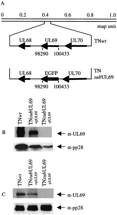
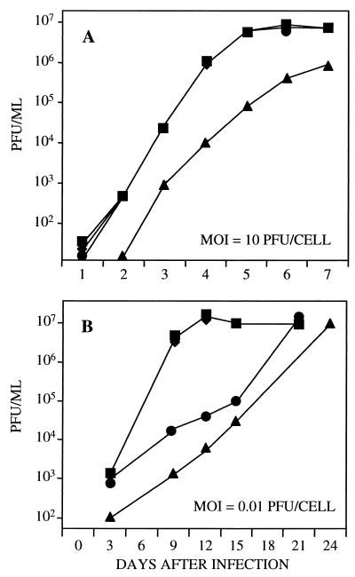
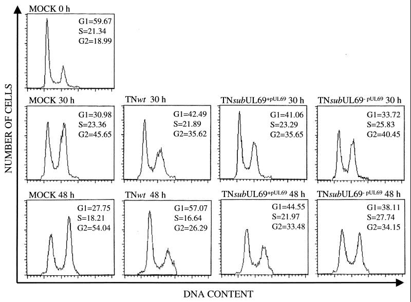
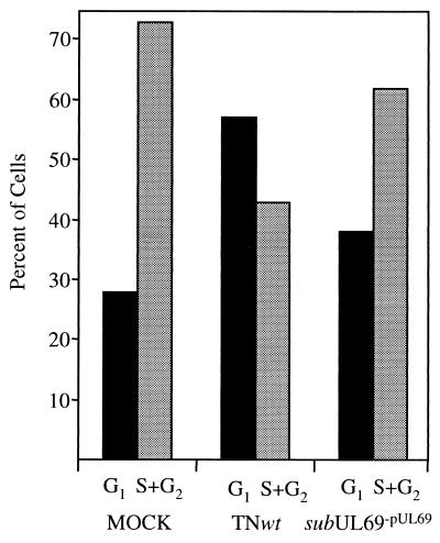
Human cytomegalovirus blocks cell-cycle progression in the G(1) compartment upon infection of primary human fibroblasts. The virus-coded UL69 protein can institute a G(1) block when expressed in cells in the absence of virus infection. We have constructed a cytomegalovirus mutant, TNsubUL69, that lacks the UL69 coding region. This virus grows slowly in fibroblasts, but produces a wild-type yield after an extended delay. It grows with normal kinetics in cells coinfected with a recombinant retrovirus, retroUL69, which expresses UL69 protein, demonstrating that its growth defect results from the mutation in the UL69 gene. UL69 protein is packaged within virus particles, and it was possible for us to produce two types of virus stocks. TNsubUL69(+pUL69) lacks the UL69 gene but contains UL69 protein in virus particles. It is produced by growth in fibroblasts that are coinfected with retroUL69. TNsubUL69(-pUL69) lacks the UL69 gene and protein. It is produced by growth in fibroblasts that do not contain UL69 protein. The mutant virions lacking both the UL69 gene and protein fail to induce a cell-cycle block with normal efficiency, whereas the mutant particles lacking the gene but containing the protein can institute the block. These results are consistent with the view that the UL69 protein contributes to the cytomegalovirus-induced cell-cycle block, and they suggest that UL69 protein delivered to cells within virions can induce the block without the synthesis of additional UL69 protein encoded by the infecting viral genome.


Figures

Figure 1
Figure 1
Construction and analysis of TNsubUL69. (A) A substitution mutant was produced in which a 2,144-bp segment within the UL69 coding region extending from sequence position 98290 to 100433 (AD169 sequence numbers) was replaced with the EGFP coding region. (B) Human fibroblasts were infected with wild-type virus (TNwt), mutant virus grown in the presence of helper retrovirus (TNsubUL69+pUL69), or mutant virus grown for one cycle in the absence of helper retrovirus (TNsubUL69−pUL69), and UL69 protein was assayed by Western blot using a UL69-specific monoclonal antibody (α-UL69) at 72 h after infection. The late protein, pp28, was assayed as a control by using a pp28-specific monoclonal antibody (α-pp28). The expression of late viral gene products is reduced in mutant virus-infected cells. (C) Virus particles were partially purified and virion proteins were assayed by Western blot.
Figure 2
Figure 2
Growth kinetics of wild-type and mutant viruses. Human fibroblasts were infected at a multiplicity of 10 pfu per cell (A) or 0.01 pfu per cell (B) with TNwt (■), TNsubUL69+pUL69 (●), TNsubUL69−pUL69 (▴), or TNsubUL69−pUL69 plus retroUL69 (♦). Cultures were harvested at the indicated times after infection, and infectious virus was assayed by plaque assay on retroUL69-infected fibroblasts. Similar results were obtained in two independent experiments.
Figure 3
Figure 3
Effect of wild-type and mutant viruses on cell-cycle progression. Human fibroblasts were mock-infected or infected at a multiplicity of 10 pfu per cell with TNwt, TNsubUL69+pUL69, or TNsubUL69−pUL69. Cultures were treated with nocodozole and phosphonoacetic acid 12 h later. Cells were harvested, fixed, and stained at either 30 or 48 h after infection, and then they were subjected to FACS analysis. The proportions of cells in G1, S, and G2 were determined by using cellquest software. As a control, mock-infected cells that were not treated with drugs (MOCK 0 h) were also analyzed.
Figure 4
Figure 4
Effects of wild-type and mutant viruses on cell-cycle progression. The bar graph compares the number of fibroblasts in G1 versus S + G2 for cultures that were either mock-infected or infected, treated with nocodozole and phosphonoacetic acid 12 h later, and then harvested and analyzed after an additional 36 h (at 48 h after infection). The primary data used to generate this graph are displayed in Fig. 3.
Figure 5
Figure 5
Accumulation of wild-type and mutant HCMV DNA. Human fibroblasts were infected at a multiplicity of 10 pfu per cell with TNwt (■), TNsubUL69+pUL69 (●), or TNsubUL69−pUL69 (▴). Cells were harvested at the indicated times after infection, viral DNA accumulation was assayed by Southern blot using 32P-labeled HCMV DNA as a probe, and radioactivity was quantified with a phosphorimager. Similar results were obtained in two independent experiments.
Figure 6
Figure 6
Accumulation of wild-type and mutant HCMV mRNA. Human fibroblasts were infected at a multiplicity of 10 pfu per cell with TNwt (white bars), TNsubUL69+pUL69 (gray bars), or TNsubUL69−pUL69 (black bars), and polyadenylated RNA was prepared at 6 h (UL123, TRS1), 24 h (UL44, UL54), or 72 h (UL82, UL83, UL99) to monitor immediate-early, early, and late mRNAs, respectively. Viral mRNA was assayed by Northern blot using 32P-labeled gene-specific probes, and radioactive bands were quantified with a phosphorimager. Similar results were obtained in two independent experiments.

References

    1. Alfred C A, Stagno S, Pass R F, Britt W. Rev Infect Dis. 1990;12:S745–S753. - PubMed
    1. Britt W, Alfred C A. In: Fields Virology. Fields B N, Knipe D M, Howley P M, editors. Philadelphia: Lippincott-Raven; 1996. pp. 2493–2523.
    1. Jault F M, Jault J M, Ruchti F, Fortunato E A, Clark C, Corbeil J, Richmann D D, Spector D H. J Virol. 1995;69:6697–6704. - PMC - PubMed
    1. Bresnahan W A, Boldogh I, Thompson E A, Albrecht T. Virology. 1996;224:150–160. - PubMed
    1. Lu M, Shenk T. J Virol. 1996;70:8850–8857. - PMC - PubMed

Publication types

LinkOut - more resources