. 2000 Apr;74(7):2981-9.
doi: 10.1128/jvi.74.7.2981-2989.2000.

Reovirus-induced apoptosis requires activation of transcription factor NF-kappaB

Affiliations

Reovirus-induced apoptosis requires activation of transcription factor NF-kappaB

J L Connolly et al. J Virol. 2000 Apr.

Abstract

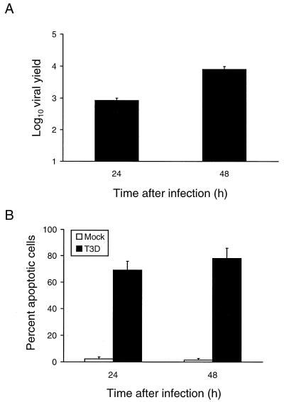
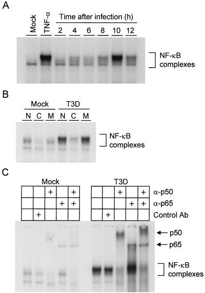
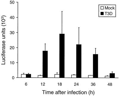
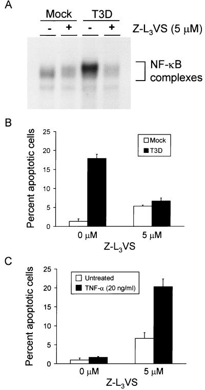
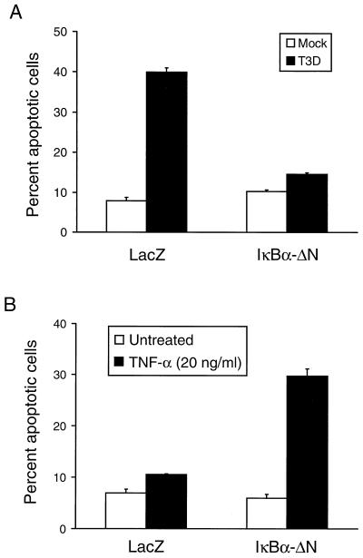
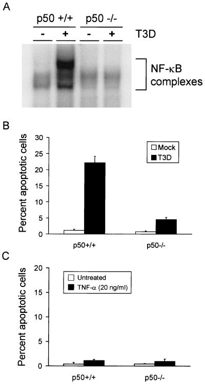
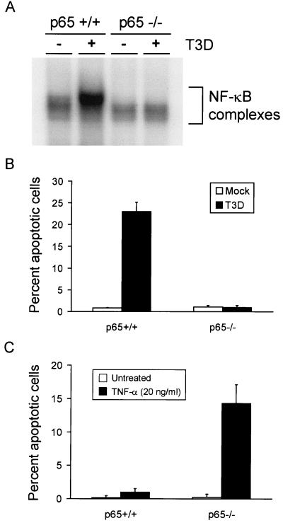
Reovirus infection induces apoptosis in cultured cells and in vivo. To identify host cell factors that mediate this response, we investigated whether reovirus infection alters the activation state of the transcription factor nuclear factor kappa B (NF-kappaB). As determined in electrophoretic mobility shift assays, reovirus infection of HeLa cells leads to nuclear translocation of NF-kappaB complexes containing Rel family members p50 and p65. Reovirus-induced activation of NF-kappaB DNA-binding activity correlated with the onset of NF-kappaB-directed transcription in reporter gene assays. Three independent lines of evidence indicate that this functional form of NF-kappaB is required for reovirus-induced apoptosis. First, treatment of reovirus-infected HeLa cells with a proteasome inhibitor prevents NF-kappaB activation following infection and substantially diminishes reovirus-induced apoptosis. Second, transient expression of a dominant-negative form of IkappaB that constitutively represses NF-kappaB activation significantly reduces levels of apoptosis triggered by reovirus infection. Third, mutant cell lines deficient for either the p50 or p65 subunits of NF-kappaB are resistant to reovirus-induced apoptosis compared with cells expressing an intact NF-kappaB signaling pathway. These findings indicate that NF-kappaB plays a significant role in the mechanism by which reovirus induces apoptosis in susceptible host cells.


Figures

FIG. 1
FIG. 1
(A) Growth of reovirus in HeLa cells. Cells (1 × 10⁵) were infected with reovirus strain T3D at an MOI of 1 PFU per cell. After adsorption for 1 h, the inoculum was removed, fresh medium was added, and the cells were incubated at 37°C for 0, 24, or 48 h. The cells were frozen and thawed twice, and viral titers were determined by a plaque assay. The results are expressed as the mean viral yields, calculated by dividing the viral titer at 24 or 48 h by the viral titer at 0 h, for three independent experiments. Error bars indicate standard error of the mean. (B) Apoptosis induced by reovirus infection of HeLa cells. Cells (5 × 10⁴) were either mock infected or infected with reovirus strain T3D at an MOI of 100 PFU per cell. After adsorption for 1 h, the cells were incubated at 37°C for 24 or 48 h and stained with acridine orange. The results are expressed as the mean percentage of cells undergoing apoptosis in three independent experiments. Error bars indicate standard error of the mean.
FIG. 2
FIG. 2
(A) Time course of NF-κB gel shift activity in nuclear extracts prepared from reovirus-infected HeLa cells. Cells (5 × 10⁶) were either mock infected or infected with T3D at an MOI of 100 PFU per cell and incubated at 37°C for the times shown. Uninfected cells also were treated with 20 ng of TNF-α per ml for 1 h. Nuclear extracts were prepared and incubated with a 32P-labeled oligonucleotide consisting of the NF-κB consensus binding sequence. Incubation mixtures were resolved by acrylamide gel electrophoresis, dried, and exposed to film. NF-κB-containing complexes are indicated. (B) Specificity of NF-κB gel shift activity. Nuclear extracts were prepared as in panel A 10 h after viral adsorption. Extracts were incubated with 32P-labeled NF-κB consensus oligonucleotide alone (lanes N), a 10-fold excess of unlabeled consensus probe (lanes C), or a 10-fold excess of unlabeled mutant probe consisting of the NF-κB consensus sequence with a point mutation that abolishes NF-κB binding (lanes M). NF-κB-containing complexes are indicated. (C) Identification of NF-κB family members activated by reovirus infection. Nuclear extracts were prepared as in panel A 10 h after viral adsorption. Extracts were incubated with no antibody, a control antibody (Ab), p50-specific antiserum, p65-specific antiserum, or both p50- and p65-specific antisera. NF-κB complexes not shifted by antibody and supershifted complexes containing p50 or p65 are indicated.
FIG. 3
FIG. 3
NF-κB-dependent luciferase expression in reovirus-infected HeLa cells. Cells (1.5 × 10⁵) were transfected with 10 μg of a luciferase reporter construct containing NF-κB binding sites. After 24 h, the cells were either mock infected or infected with T3D at an MOI of 100 PFU per cell and incubated at 37°C for the times shown. Cell extracts were prepared, and luciferase activity was determined. The results are expressed as the mean luciferase units in two independent experiments. Error bars indicate standard error of the mean.
FIG. 4
FIG. 4
(A) NF-κB gel shift activity in reovirus-infected cells cultured in the presence of Z-L3VS. Cells (5 × 10⁶) were either mock infected or infected with T3D at an MOI of 100 PFU per cell and cultured in the absence or presence of 5 μM Z-L3VS. After incubation at 37°C for 10 h, nuclear extracts were prepared and incubated with a 32P-labeled oligonucleotide consisting of the NF-κB consensus sequence. Incubation mixtures were resolved by acrylamide gel electrophoresis, dried, and exposed to film. NF-κB-containing complexes are indicated. (B) Quantitation of apoptosis in reovirus-infected HeLa cells cultured in the presence of Z-L3VS. Cells (5 × 10⁴) were either mock infected or infected with T3D at an MOI of 100 PFU per cell and cultured in the absence or presence of 5 μM Z-L3VS. After incubation at 37°C for 18 h, the cells were stained with acridine orange. (C) Quantitation of apoptosis in TNF-α-treated HeLa cells cultured in the presence of Z-L3VS. Cells (5 × 10⁴) were either untreated or treated with 20 ng of TNF-α per ml and cultured in the absence or presence of 5 μM Z-L3VS. After incubation at 37°C for 18 h, the cells were stained with acridine orange. The results of the experiments in panels B and C are expressed as the mean percentage of cells undergoing apoptosis in three independent experiments. Error bars indicate standard error of the mean.
FIG. 5
FIG. 5
Quantitation of apoptosis in HeLa cells transiently expressing IκBα-ΔN. Cells (1 × 10⁶) were either transfected with 5 μg of pHook-2/IκBα-ΔN or 5 μg of pHook-2/lacZ. After 24 h, transfected cells were isolated using Capture-Tec magnetic beads and plated in 24-well plates. (A) Transfected cells (2 × 10⁴) were either mock infected or infected with T3D at an MOI of 100 PFU per cell. After incubation at 37°C for 24 h, the cells were stained with acridine orange. (B) Transfected cells (2 × 10⁴) were either not treated or treated with 20 ng of TNF-α per ml. After incubation at 37°C for 24 h, the cells were stained with acridine orange. The results of the experiments in both panels are expressed as the mean percentage of cells undergoing apoptosis in three independent experiments. Error bars indicate standard error of the mean.
FIG. 6
FIG. 6
(A) NF-κB gel shift activity in reovirus-infected p50+/+ and p50−/− immortalized fibroblast cells. Cells (5 × 10⁶) were either mock infected or infected with T3D at an MOI of 100 PFU per cell. After incubation at 37°C for 6 h, nuclear extracts were prepared and incubated with a 32P-labeled DNA probe consisting of the NF-κB consensus sequence. Incubation mixtures were resolved by acrylamide gel electrophoresis, dried, and exposed to film. NF-κB-containing complexes are indicated. (B) Quantitation of apoptosis in reovirus-infected p50+/+ and p50−/− cells. Cells (2.5 × 10⁴) were either mock infected or infected with T3D at an MOI of 100 PFU per cell. After incubation at 37°C for 48 h, the cells were stained with acridine orange. (C) Quantitation of apoptosis in TNF-α-treated p50+/+ and p50−/− cells. Cells (2.5 × 10⁴) were either untreated or treated with 20 ng of TNF-α per ml. After incubation at 37°C for 24 h, the cells were stained with acridine orange. The results of the experiments in panels B and C are expressed as the mean percentage of cells undergoing apoptosis in three independent experiments. Error bars indicate standard error of the mean.
FIG. 7
FIG. 7
(A) NF-κB gel shift activity in reovirus-infected p65+/+ and p65−/− immortalized fibroblast cells. Cells (5 × 10⁶) were either mock infected or infected with T3D at an MOI of 100 PFU per cell. After incubation at 37°C for 8 h, nuclear extracts were prepared and incubated with a 32P-labeled DNA probe consisting of the NF-κB consensus sequence. Incubation mixtures were resolved by acrylamide gel electrophoresis, dried, and exposed to film. NF-κB-containing complexes are indicated. (B) Quantitation of apoptosis in reovirus-infected p65+/+ and p65−/− cells. Cells (2.5 × 10⁴) were either mock infected or infected with T3D at an MOI of 100 PFU per cell. After incubation at 37°C for 48 h, the cells were stained with acridine orange. (C) Quantitation of apoptosis in TNF-α-treated p65+/+ and p65−/− cells. Cells (2.5 × 10⁴) were either untreated or treated with 20 ng of TNF-α per ml. After incubation at 37°C for 24 h, the cells were stained with acridine orange. The results of the experiments in panels B and C are expressed as the mean percentage of cells undergoing apoptosis in three independent experiments. Error bars indicate standard error of the mean.
FIG. 8
FIG. 8
Growth of reovirus in p50−/− and p65−/− immortalized fibroblast cells. p50+/+ and p50−/− cells (A) or p65+/+ and p65−/− cells (B) (2.5 × 10⁴ cells per experiment) were infected with T3D at an MOI of 1 PFU per cell. After adsorption for 1 h, the inoculum was removed, fresh medium was added, and the cells were incubated at 37°C for 0, 24, or 48 h. The cells were frozen and thawed twice, and viral titers were determined by a plaque assay. The results are presented as the mean viral yields (viral titer at 24 or 48 h divided by viral titer at 0 h) in three independent experiments. Error bars indicate standard error of the mean.

Similar articles

Cited by

References

    1. Abbadie C, Kabrun N, Bouali F, Vandenbunder B, Enrietto P. High levels of c-rel expression are associated with programmed cell death in the developing avian embryo and in bone marrow cells in vitro. Cell. 1993;75:899–912. - PubMed
    1. Ashkenazi A, Dixit V M. Death receptors: signaling and modulation. Science. 1998;281:1305–1308. - PubMed
    1. Baeuerle P, Baltimore D. IκB: a specific inhibitor of the NF-κB transcription factor. Science. 1988;242:540–546. - PubMed
    1. Baeuerle P, Baltimore D. A 65-kD subunit of active NF-κB is required for inhibition of NF-κB by IκB. Genes Dev. 1989;3:1689–1698. - PubMed
    1. Ballard D W, Bohnlein E, Lowenthal J W, Wano Y, Franza B R, Greene W C. HTLV-I tax induces cellular proteins that activate the kappa B element in the IL-2 receptor alpha gene. Science. 1988;241:1652–1655. - PubMed

Publication types