. 2000 Apr;74(7):3046-57.
doi: 10.1128/jvi.74.7.3046-3057.2000.

Basic residues in human immunodeficiency virus type 1 nucleocapsid promote virion assembly via interaction with RNA

Affiliations

Basic residues in human immunodeficiency virus type 1 nucleocapsid promote virion assembly via interaction with RNA

A Cimarelli et al. J Virol. 2000 Apr.

Abstract

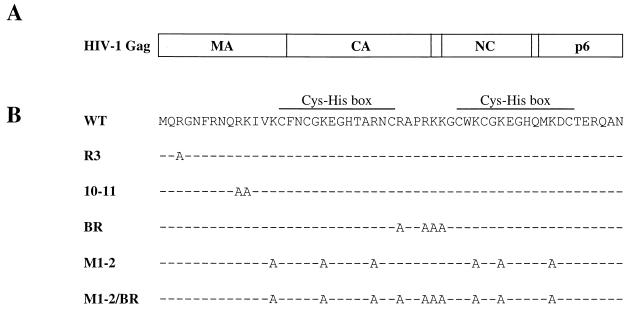
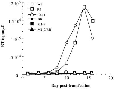
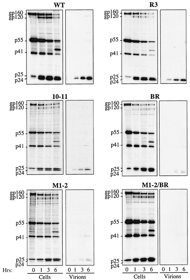
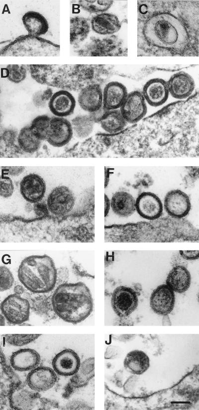
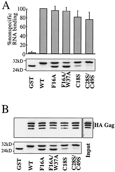
Retroviral Gag polyproteins drive virion assembly by polymerizing to form a spherical shell that lines the inner membrane of nascent virions. Deletion of the nucleocapsid (NC) domain of the Gag polyprotein disrupts assembly, presumably because NC is required for polymerization. Human immunodeficiency virus type 1 NC possesses two zinc finger motifs that are required for specific recognition and packaging of viral genomic RNA. Though essential, zinc fingers and genomic RNA are not required for virion assembly. NC promiscuously associates with cellular RNAs, many of which are incorporated into virions. It has been hypothesized that Gag polymerization and virion assembly are promoted by nonspecific interaction of NC with RNA. Consistent with this model, we found an inverse relationship between the number of NC basic residues replaced with alanine and NC's nonspecific RNA-binding activity, Gag's ability to polymerize in vitro and in vivo, and Gag's capacity to assemble virions. In contrast, mutation of NC's zinc fingers had only minor effects on these properties.


Figures

FIG. 1
FIG. 1
(A) Schematic representation of the HIV-1 Gag polyprotein showing the major domains. Vertical lines, positions of viral protease cleavage sites. (B) Amino acid sequence of HIV-1 NC. The sequences of the mutants used in this study are shown. The names of the mutant NC proteins are on the left. Dashes indicate amino acid residues identical to those of the wild type (WT).
FIG. 2
FIG. 2
HIV-1 replication kinetics following transfection of wild-type (WT) or NC mutant proviral DNAs into the Jurkat T-cell line. The accumulation of RT activity in the cell culture supernatant is shown for the indicated day posttransfection.
FIG. 3
FIG. 3
Pulse-chase analysis of HIV-1 NC mutants. HeLa cells transfected with the indicated NL4-3 proviral DNAs were metabolically labeled with [35S]Met-[35S]Cys for 45 min and chased with unlabeled media for 0, 1, 3, and 6 h, as indicated. Virion-associated proteins were purified by ultracentrifugation through 25% sucrose. Virion- and cell-associated proteins were immunoprecipitated using sera from an HIV-1-infected individual and analyzed by SDS-PAGE. The mobilities of the envelope glycoprotein precursor (gp160), surface envelope protein (gp120), Pr55Gag precursor (p55), incompletely processed Gag precursors (p41 and p25), and completely processed CA (p24) are indicated on the left. WT, wild type.
FIG. 4
FIG. 4
Analysis of HIV-1 NC mutant virion morphology. 293T cells were transfected with proviral DNAs, either wild type (A and B), mutant R3 (C and D), mutant 10-11 (E and F), mutant BR (G and H), mutant M1-2 (I), or mutant M1-2/BR (J). Cells were fixed, stained, embedded, and visualized by electron microscopy. Bar, 100 nm.
FIG. 5
FIG. 5
Determination of HIV-1 NC mutant virion density. Virions produced by transfection of the indicated proviral DNAs into 293T cells were purified, concentrated, and layered onto a linear sucrose gradient (20 to 60%). Eleven fractions (abscissa) were collected from the top of the gradient. Left ordinate, fraction density; right ordinate, exogenous RT activity in each fraction.
FIG. 6
FIG. 6
RNA-binding activity of HIV-1 NC mutants. (A) Twofold dilutions of purified wild-type HIV-1 virions were loaded onto a nylon membrane and probed with a 32P-labeled DNA oligonucleotide specific for viral genomic RNA. (B) Wild-type (WT) and NC mutant virions were purified, normalized by exogenous RT activity, and subjected to dot blot analysis as for panel A. Results are presented as percentages of wild-type virus activity. The graph presents means from three independent experiments with standard errors of the means. Primary data from a representative experiment are shown underneath. Mock, “virion preparation” from cells transfected with a myristylation-deficient NL4-3. (C) GST protein fused to wild-type NC was expressed in bacteria. After serial twofold dilutions, the protein was purified using glutathione-agarose beads and bound in solution to 32P-labeled, nonspecific RNA. After a washing, RNA that remained bound was quantitated directly in a beta counter. Bound proteins were visualized by Coomassie staining after SDS-PAGE (bottom). (D) NC basic-residue mutants were expressed as GST fusion proteins and analyzed as described for panel C. RNA-binding activity is indicated as a percentage of the wild type. The graph presents means from three independent experiments with standard errors of the means. The bound fusion proteins were visualized by Coomassie staining after SDS-PAGE (bottom).
FIG. 7
FIG. 7
NC basic-residue mutants disrupt Gag-Gag interaction. (A) NC mutants were expressed as GST fusion proteins in bacteria, immobilized onto glutathione-agarose beads and incubated with a lysate from bacteria expressing HA-Gag. After a washing, proteins bound to the beads were boiled, processed by SDS-PAGE, and analyzed by Western blotting (WB) with an anti-HA antibody (top) or by Coomassie staining (bottom). The input lane shows 10% of the total HA-Gag bacterial lysate used in the binding reactions. The positions of migration of molecular mass markers (in kilodaltons) are indicated on the left of the Coomassie gel. WT, wild type. (B) Schematic representation of in vivo complementation assay. Expression of protease-defective HIV-1 provirus (white rectangle) by transfection of 293T cells leads to the release of immature particles in the supernatant. HA-Gag (gray rectangle) produced by transfection does not release particles because this protein is not myristylated. Coexpression of protease-defective HIV-1 and HA-Gag in the same cell produces mixed particles if HA-Gag is rescued via interaction with the provirally encoded Gag. (C) HA-Gags, wild type or bearing the NC mutations indicated, were coexpressed with protease-defective HIV-1 provirus. Particles purified from the supernatant were normalized by exogenous RT and analyzed by Western blotting using anti-HA (top) or anti-cyclophilin A (middle) antibodies. Transfected cell lysates were analyzed by Western blotting using anti-HA (bottom). A Gag polyprotein with a deletion of the NC and p6 domains (ΔNC-p6) was included as a negative control. The positions of migration of the proteins are on the right.
FIG. 8
FIG. 8
NC basic residues are required for the formation of intracellular, detergent-resistant Gag complexes. HeLa cells were transfected with either wild-type or M1-2/BR-bearing proviruses as indicated. Proteins were metabolically labeled for 2 h, and cells were lysed in 1% Triton X-100. The cytoplasmic fraction was loaded onto a linear sucrose gradient (20 to 60%) and accelerated at 80,000 × g for 24 h. Thirteen fractions were harvested from top to bottom as indicated. The solution density of each fraction was measured (A). Viral proteins were immunoprecipitated, processed by SDS-PAGE, and detected with a phosphorimager (B).
FIG. 9
FIG. 9
Analysis of HIV-1 NC Cys-His box mutants. (A) The indicated HIV-1 NC Cys-His box mutants were expressed as GST fusion proteins in bacteria and immobilized onto glutathione-agarose beads. Binding of 32P-labeled RNA was assessed as described for Fig. 6. The graph presents means from three independent experiments with standard errors of the means. (B) GST-NC proteins bearing Cys-His box mutants were assayed for the ability to bind full-length HA-tagged Gag in vitro, as described in the legend of Fig. 7A. Samples were analyzed by Western blotting with an anti-HA antibody (top) or by Coomassie staining (bottom). The input lane shows 10% of the total HA-Gag bacterial lysate used in the binding reaction. The positions of migration of molecular mass markers (in kilodaltons) are indicated on the left of the Coomassie gel.

References

    1. Aldovini A, Young R. Mutations of RNA and protein sequences involved in human immunodeficiency virus type 1 packaging result in production of noninfectious virus. J Virol. 1990;64:1920–1926. - PMC - PubMed
    1. Bennett R P, Nelle T D, Wills J W. Functional chimeras of the Rous sarcoma virus and human immunodeficiency virus Gag proteins. J Virol. 1993;67:6487–6498. - PMC - PubMed
    1. Berkowitz R, Fisher J, Goff S P. RNA packaging. Curr Top Microbiol Immunol. 1996;214:177–218. - PubMed
    1. Berthoux L, Pechoux C, Ottmann M, Morel G, Darlix J L. Mutations in the N-terminal domain of human immunodeficiency virus type 1 nucleocapsid protein affect virion core structure and proviral DNA synthesis. J Virol. 1997;71:6973–6981. - PMC - PubMed
    1. Bowzard J B, Bennett R P, Krishna N K, Ernst S M, Rein A, Wills J W. Importance of basic residues in the nucleocapsid sequence for retrovirus Gag assembly and complementation rescue. J Virol. 1998;72:9034–9044. - PMC - PubMed

Publication types

MeSH terms

LinkOut - more resources