. 2000 Apr;74(7):3093-104.
doi: 10.1128/jvi.74.7.3093-3104.2000.

A protein kinase activity associated with Epstein-Barr virus BGLF4 phosphorylates the viral early antigen EA-D in vitro

Affiliations

A protein kinase activity associated with Epstein-Barr virus BGLF4 phosphorylates the viral early antigen EA-D in vitro

M R Chen et al. J Virol. 2000 Apr.

Abstract

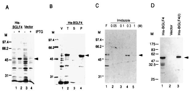
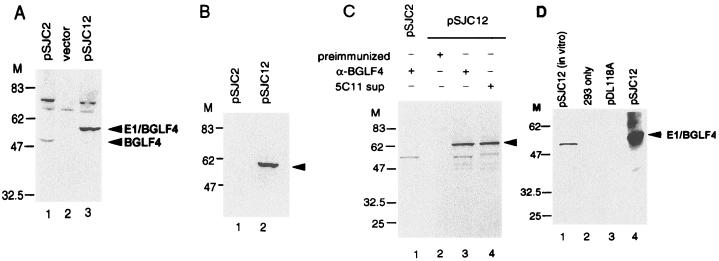
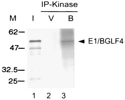
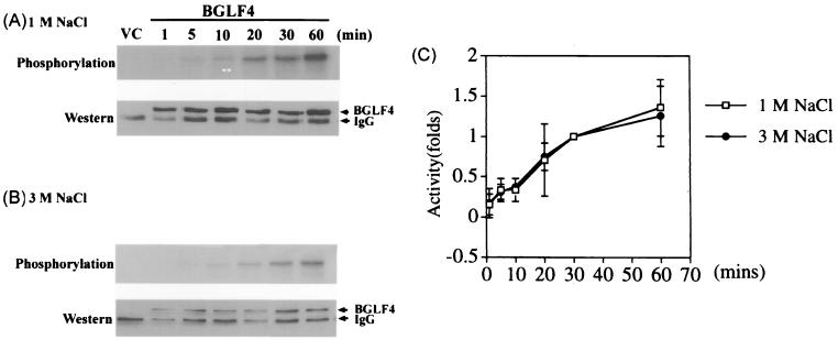
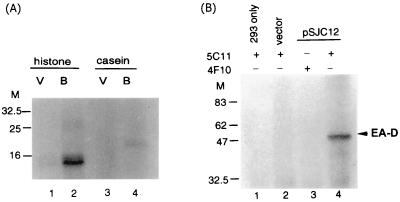
The Epstein-Barr virus (EBV) open reading frame BGLF4 was identified as a potential Ser/Thr protein kinase gene through the recognition of amino acid sequence motifs characteristic of conserved regions within the catalytic domains of protein kinases. In order to investigate this potential kinase activity, BGLF4 was expressed in Escherichia coli and the purified protein was used to generate a specific antiserum. Recombinant vaccinia virus vTF7-3, which expresses the T7 RNA polymerase, was used to infect 293 and 293T cells after transient transfection with a plasmid containing BGLF4 under the control of the T7 promoter. Autophosphorylation of the BGLF4 protein was demonstrated using the specific antiserum in an immune complex kinase assay. In addition, EBNA-1-tagged BGLF4 and EBNA-1 monoclonal antibody 5C11 were used to demonstrate the specificity of the kinase activity and to locate BGLF4 in the cytoplasm of transfected cells. Manganese ions were found to be essential for autophosphorylation of BGLF4, and magnesium can stimulate the activity. BGLF4 can utilize GTP, in addition to ATP, as a phosphate donor in this assay. BGLF4 can phosphorylate histone and casein in vitro. Among the potential viral protein substrates we examined, the EBV early antigen (EA-D, BMRF1), a DNA polymerase accessory factor and an important transactivator during lytic infection, was found to be phosphorylated by BGLF4 in vitro. Amino acids 1 to 26 of BGLF4, but not the predicted conserved catalytic domain, were found to be essential for autophosphorylation of BGLF4.


Figures

FIG. 1
FIG. 1
Expression of His-BGLF4 in E. coli BL21(DE3) and generation of BGLF4-specific antiserum. (A) Total cell lysate from bacteria carrying vector control (pRSETA) or pSJC1 (His-BGLF4). Lysate is shown uninduced (lanes 1 and 3) and after induction with IPTG (lanes 2 and 4) and was displayed on a 10% PAGE gel and stained with Coomassie blue. The predicted molecular mass of BGLF4 is approximately 52 kDa, as indicated by the arrowhead. (B) After lysis with buffer (lane 2; total lysate), the cell lysate was fractionated into soluble proteins (lane 3) and pellet. Most of the BGLF4 product appeared in insoluble form in E. coli (lane 4). V, vector control. (C) The recombinant BGLF4 protein was dissolved in 8 M urea–5 mM imidazole–100 mM NaCl and purified using a nickel column. After being washed with binding buffer (lane 2) and 100 mM imidazole buffer (lane 3), the BGLF4 protein was eluted with buffer containing 300 mM imidazole (lane 4). F, flowthrough. (D) Western immunoblotting demonstrates the specificity of rabbit anti-BGLF4 antiserum. Total cell lysates of E. coli carrying pSJC1 (lane 1) or pRSETA (lane 2) and purified BGLF4 (lane 3) were displayed on an SDS–10% PAGE gel, transferred onto a membrane, and probed with rabbit anti-BGLF4 antiserum as described in Materials and Methods. M, molecular weight markers.
FIG. 2
FIG. 2
Expression of BGLF4 (pSJC2) and E1/BGLF4 (pSJC12) in 293 cells after transfection and infection with recombinant vaccinia virus vTF7-3, which carries a copy of the T7 RNA polymerase. (A) After transfection and infection, cell lysates of pSG5 (vector), pSJC2(pSG5-BGLF4), and pSJC12(pSG5-E1/BGLF4) were harvested, displayed on an SDS–10% PAGE gel, and reacted with BGLF4-specific antiserum in an immunoblotting assay. The 48-kDa product of BGLF4 and the 52-kDa product of E1/BGLF4 can be seen in lanes 1 and 3, respectively. (B) The cell lysates expressing BGLF4 and E1/BGLF4 were immunoblotted with EBNA-1 monoclonal antibody 5C11. Arrowhead, E1/BGLF4. (C) After transfection of BGLF4 or E1/BGLF4 expression plasmids and infection with vTF7-3, the cells were labeled with [35S]methionine for 4 h before lysis. The cell lysates were immunoprecipitated with preimmunized-rabbit sera, BGLF4-specific antisera, or the 5C11 monoclonal antibody. Arrowhead, E1/BGLF4. (D) Autophosphorylation of BGLF4. Protein A-Sepharose beads containing immunoprecipitated E1/BGLF4 were incubated in kinase buffer in the presence of [γ-32P]ATP. Autophosphorylation of BGLF4 may be seen in lane 4. Lane 1, in vitro transcription/translation product as a marker.
FIG. 3
FIG. 3
Optimal conditions for BGLF4 autophosphorylation. Protein A-Sepharose beads containing immunoprecipitated E1/BGLF4 were incubated in various kinase buffers in the presence of [γ-32P]ATP. (A) Effect of pH on BGLF4 kinase activity. The buffer containing 1 mM MnCl2, 1 mM MgCl2, 150 mM NaCl, 0.5% NP-40, and 50 mM HEPES was adjusted to different pHs as indicated. The autophosphorylation activities are similar in the range of pH 6.5 to 8.0, whereas the activities are lower at pH 6.2 and pH 8.3. (B) Effects of divalent cations on BGLF4 kinase activity. The buffer containing 50 mM HEPES (pH 7.4), 150 mM NaCl, and various amounts of MgCl2 and MnCl2, as indicated. Manganese is essential for BGLF4 activity (lane 6), which can be further stimulated by magnesium (lane 5). (C) Effects of monovalent cations on BGLF4 kinase activity. The buffer contained 50 mM HEPES (pH 7.4), 10 mM MgCl2, and 10 mM MnCl2, as indicated, and different concentrations of NaCl or KCl. The maximal activity was observed in the presence of 300 mM KCl (lane 6). (D) Effect of detergent on the BGLF4 kinase activity. In addition to 50 mM HEPES (pH 7.4), 10 mM MgCl2, 10 mM MnCl2, and 300 mM KCl, 0.5% NP-40 appeared to stimulate BGLF4 kinase activity. The in vitro transcription/translation product of BGLF4 (I) is shown as a marker. The immunoprecipitation product of pDL118-transfected cell lysate was also used in the kinase assay as a negative control (V) in each panel.
FIG. 4
FIG. 4
Utilization of GTP as phosphate donor for BGLF4 autophosphorylation. The immunoprecipitation product of E1/BGLF4, as described for Fig. 3, was used for autophosphorylation in the kinase buffer containing [γ-32P]GTP instead of [γ-32P]ATP (lane 3). I, [35S]methionine-labeled in vitro transcription/translation product; V, vector (pPDL118) control.
FIG. 5
FIG. 5
Kinetics of immunoprecipitated BGLF4 autophosphorylation using buffer containing 1 or 3 M NaCl to wash the immunocomplexes. Cell lysate (30 μg) harvested from pSJC12-transfected and vTF7-3-infected 293T cells was used for immunoprecipitation. The immunocomplexes were washed with 1 or 3 M NaCl buffer and incubated with [γ-32P]ATP in kinase buffer for various periods of time, as indicated. The reaction mixture containing vector control (VC) was incubated for 30 min. The products were displayed on SDS–10% PAGE gel, transferred to Hybond-C membranes, and quantitated by phosphorimager (A and B, top). The membranes were immunoblotted with 5C11 and developed with an ECL kit to demonstrate the relative amounts of BGLF4 in each reaction mixture (A and B, bottom), and the exposed films were scanned and quantitated with the Scion Imager (National Institutes of Health) program. (C) The kinase activities of the reaction mixtures were divided by the relative amount of protein in each reaction mixture, and the averages of three independent experiments were indicated as relative fold increases or decreases. The kinase activity at 30 min was counted as 1.
FIG. 6
FIG. 6
Autophosphorylation of BGLF4 was not affected in the presence of heparin or okadaic acid. (A) Different concentrations of heparin, as indicated, were included in the autophosphorylation reaction mixture for E1/BGLF4 (lanes 2 to 6). Ten units of CK II phosphorylated 1 μg of casein, and this phosphorylation was completely blocked in the presence of heparin (lanes 7 and 8). VC, vector control. (D) The addition of phosphatase inhibitor okadaic acid (OA) did not increase the autophosphorylation of BGLF4. (B and E) The same blots as in panels A and D were probed with 5C11 to show the relative amounts of E1/BGLF4 in the reaction mixtures. (C and F) The kinase activities of each reaction mixture were divided by the relative amounts of protein in each reaction mixture, and the averages of three independent experiments were indicated as relative fold increases or decreases.
FIG. 7
FIG. 7
Analysis of the phosphoamino acids of BGLF4, using phosphoamino acid-specific monoclonal antibodies. After the immune complex kinase assay, the products were displayed on SDS–10% PAGE gel, transferred onto Hybond-C membranes, and immunoblotted with antiphosphoserine, antiphosphothreonine, or antiphosphotyrosine specific monoclonal antibodies. Arrowheads, positions of BGLF4 and immunoglobulin G (IgG). VC, vector control.
FIG. 8
FIG. 8
BGLF4 expressed in the cytoplasm of pCF4-transfected 293T cells. (A) 293T cells transfected with pCF4 were fixed with 50% methanol–50% acetone and reacted with 5C11 monoclonal antibody. (B) Photograph of cells reacted with anti-BGLF4 under lower magnification.
FIG. 9
FIG. 9
Transphosphorylation of BGLF4. Protein A-Sepharose beads containing immunoprecipitated E1/BGLF4 were incubated with 1 μg of individual protein in the presence of 5 μCi of [γ-32P]ATP. After incubation, the products were precipitated with TCA and analyzed on an SDS–10% PAGE gel. (A) A stronger signal was observed in the reaction of histone (lane 2) than in that of casein (lane 4). V, cell lysate from vector control; B, cell lysate from E1/BGLF4-transfected cells. (B) Purified bacterially expressed EA-D was also phosphorylated by the 5C11-immunoprecipitated E1/BGLF4 product. 4F10 is a monoclonal antibody against EBV DNase which was used as a negative control in immunoprecipitation.
FIG. 10
FIG. 10
(A) Sequence alignment of BGLF4 and its homologues among other herpesviruses using the CLUSTALW program (25, 53). Amino acids conserved among herpesviruses relative to the N terminus at amino acid 231 of BGLF4 are shaded. Conserved boxes I to IV are underlined; ∗, amino acids chosen for mutation. (B) Summary of BGLF4 mutants and their relative activities. The BGLF4 coding region encodes 429 amino acids. The putative functional domains, based on alignment of different kinases, are indicated at the top. Different mutants were generated by PCR or recombinant PCR as described in Materials and Methods. Amino acid changes to methionine (M) or alanine (A) are indicated. Relative activities of autophosphorylation are summarized on the right.
FIG. 11
FIG. 11
Relative autophosphorylation activities of BGLF4 mutants. All the plasmids were expressed in 293T cells by transfection and infection of vTF7-3 and assayed for autophosphorylation activities as described for Fig. 6. (A) The immune complex kinase products were displayed on SDS–12% PAGE gel and transferred to a Hybond-C membrane and exposed to X-ray film. The autophosphorylation level was quantitated by STORM 842. (B) The Hybond-C membrane was then probed with the 5C11 monoclonal antibody, and the specific bands were quantitated with the Scion Imager program. (C) The kinase activities of individual lanes were divided by the relative amount of protein in each reaction mixture, and the averages of three independent experiments are indicated as relative fold increases or decreases.
FIG. 12
FIG. 12
The sequence of the first 26 amino acids of BGLF4 contains multiple serine residues and a possible glycosylation site (underlined).

References

    1. Ali M A, Prakash S S, Jariwalla R. Localization of the antigenic sites and intrinsic protein kinase domain within a 300 amino acid segment of the ribonucleotide reductase large subunit from herpes simplex virus type 2. Virology. 1992;187:360–367. - PubMed
    1. Beaud G, Sharif A, Topa-Masse A, Leader D P. Ribosomal protein S2/Sa kinase purified from HeLa cells infected with vaccinia virus corresponds to the B1R protein kinase and phosphorylates in vitro the viral ssDNA-binding protein. J Gen Virol. 1994;75:283–293. - PubMed
    1. Branch D R, Mills G B. pp60c-src expression is induced by activation of normal human T lymphocytes. J Immunol. 1995;154:3678–3685. - PubMed
    1. Cannon J S, Hamzeh F, Moore S, Nicholas J, Ambinder R F. Human herpesvirus 8-encoded thymidine kinase and phosphotransferase homologues confer sensitivity to ganciclovir. J Virol. 1999;73:4786–4793. - PMC - PubMed
    1. Chee M S, Lawrence G L, Barrell B G. Alpha-, beta- beta- and gammaherpesviruses encode a putative phosphotransferase. J Gen Virol. 1989;70:1151–1160. - PubMed

Publication types

MeSH terms

LinkOut - more resources