. 2000 Apr;74(7):3188-95.
doi: 10.1128/jvi.74.7.3188-3195.2000.

A recombinant human parainfluenza virus type 3 (PIV3) in which the nucleocapsid N protein has been replaced by that of bovine PIV3 is attenuated in primates

Affiliations

A recombinant human parainfluenza virus type 3 (PIV3) in which the nucleocapsid N protein has been replaced by that of bovine PIV3 is attenuated in primates

J E Bailly et al. J Virol. 2000 Apr.

Abstract

The shipping fever (SF) and Kansas (Ka) strains of bovine parainfluenza virus type 3 (BPIV3) are restricted in their replication in rhesus monkeys 100- to 1,000-fold compared to human parainfluenza virus type 3 (HPIV3), and the Ka strain also was shown to be attenuated in humans. To initiate an investigation of the genetic basis of the attenuation of BPIV3 in primates, we produced viable chimeric HPIV3 recombinants containing the nucleoprotein (N) open reading frame (ORF) from either BPIV3 Ka or SF in place of the HPIV3 N ORF. These chimeric recombinants were designated cKa-N and cSF-N, respectively. Remarkably, cKa-N and cSF-N grew to titers comparable to those of their HPIV3 and BPIV3 parents in LLC-MK2 monkey kidney and Madin-Darby bovine kidney cells. Thus, the heterologous nature of the N protein did not impede replication in vitro. However, cKa-N and cSF-N were each restricted in replication in rhesus monkeys to a similar extent as Ka and SF, respectively. This identified the BPIV3 N protein as a determinant of the host range restriction of BPIV3 in primates. These chimeras thus combine the antigenic determinants of HPIV3 with the host range restriction and attenuation phenotype of BPIV3. Despite their restricted replication in rhesus monkeys, the chimeric viruses induced a level of resistance to HPIV3 challenge in these animals which was indistinguishable from that conferred by immunization with HPIV3. The infectivity, attenuation, and immunogenicity of these BPIV3/HPIV3 chimeras suggest that the modified Jennerian approach described in the present report represents a novel method to design vaccines to protect against HPIV3-induced disease in humans.


Figures

FIG. 1
FIG. 1
Features of the BPIV3/HPIV3 chimeric genomes and their confirmation by TaqI digestion of RT-PCR products generated from vRNA. (A) The genomes of the chimeric cKa-N and cSF-N viruses are shown schematically (not to scale) relative to those of the HPIV3 and BPIV3 parents. Arrows above the rJS genome indicate the locations of primers used for RT-PCR amplification of chimeric and parent viruses for diagnostic TaqI digestion and sequence analysis. These primers were directed to regions conserved between HPIV3 and BPIV3 so that they could be used for the amplification of HPIV3, BPIV3, and BPIV3/HPIV3 chimeras. (B) Expected sizes of TaqI digestion products for each virus are shown. TaqI fragments which are unique to each virus and therefore serve in virus identification are indicated by stars. (C) TaqI profiles of RT-PCR products containing the PIV3 N coding region of chimeric cKa-N (left) and cSF-N (right) are shown flanked by those of the HPIV3 and BPIV3 parents. RT-PCR products (1.9 kb) containing the PIV3 N coding region and flanking sequence were amplified from virion RNA using primers whose locations are shown in (panel A) and subjected to digestion with TaqI. Unique TaqI fragments diagnostic of virus identity and corresponding to those identified in panel B are indicated by stars. Calculated lengths of DNA gel bands are indicated.
FIG. 1
FIG. 1
Features of the BPIV3/HPIV3 chimeric genomes and their confirmation by TaqI digestion of RT-PCR products generated from vRNA. (A) The genomes of the chimeric cKa-N and cSF-N viruses are shown schematically (not to scale) relative to those of the HPIV3 and BPIV3 parents. Arrows above the rJS genome indicate the locations of primers used for RT-PCR amplification of chimeric and parent viruses for diagnostic TaqI digestion and sequence analysis. These primers were directed to regions conserved between HPIV3 and BPIV3 so that they could be used for the amplification of HPIV3, BPIV3, and BPIV3/HPIV3 chimeras. (B) Expected sizes of TaqI digestion products for each virus are shown. TaqI fragments which are unique to each virus and therefore serve in virus identification are indicated by stars. (C) TaqI profiles of RT-PCR products containing the PIV3 N coding region of chimeric cKa-N (left) and cSF-N (right) are shown flanked by those of the HPIV3 and BPIV3 parents. RT-PCR products (1.9 kb) containing the PIV3 N coding region and flanking sequence were amplified from virion RNA using primers whose locations are shown in (panel A) and subjected to digestion with TaqI. Unique TaqI fragments diagnostic of virus identity and corresponding to those identified in panel B are indicated by stars. Calculated lengths of DNA gel bands are indicated.
FIG. 2
FIG. 2
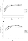
Multicycle growth of parental and chimeric viruses in MDBK (A) or LLC-MK2 (B) cells. Monolayers of MDBK (A) or LLC-MK2 (B) cells in wells (9.6 cm2 each) of a six-well plate were infected at an MOI of 0.01 with the indicated parental or chimeric virus. Three replicate infections were performed with each virus. Samples were taken at the indicated time points and stored at −70°C, and the TCID50 of each sample was determined in one assay. Growth curves were constructed using the average of three replicate samples at each time point. The lower limit of virus detectability was 101.5 TCID50/ml, which is indicated by a horizontal dotted line.

Similar articles

Cited by

References

    1. Bailly, J. E., J. M. McAuliffe, M. H. Skiadopoulos, P. L. Collins, and B. R. Murphy. Sequence determination and molecular analysis of two strains of bovine parainfluenza virus type 3 that are attenuated for primates. Virus Genes, in press. - PubMed
    1. Belshe R B, Hissom F K. Cold adaptation of parainfluenza virus type 3: induction of three phenotypic markers. J Med Virol. 1982;10:235–242. - PubMed
    1. Clements M L, Belshe R B, King J, Newman F, Westblom T U, Tierney E L, London W T, Murphy B R. Evaluation of bovine, cold-adapted human, and wild-type human parainfluenza type 3 viruses in adult volunteers and in chimpanzees. J Clin Microbiol. 1991;29:1175–1182. - PMC - PubMed
    1. Clements M L, Sears S D, Christina K, Murphy B R, Snyder M H. Comparison of the virologic and immunologic responses of volunteers to live avian-human influenza A H3N2 reassortant virus vaccines derived from two different avian influenza virus donors. J Clin Microbiol. 1989;27:219–222. - PMC - PubMed
    1. Clements M L, Subbarao E K, Fries L F, Karron R A, London W T, Murphy B R. Use of single-gene reassortant viruses to study the role of avian influenza A virus genes in attenuation of wild-type human influenza A virus for squirrel monkeys and adult human volunteers. J Clin Microbiol. 1992;30:655–662. - PMC - PubMed

LinkOut - more resources