. 2000 Apr;74(7):3418-22.
doi: 10.1128/jvi.74.7.3418-3422.2000.

Assembly and processing of human immunodeficiency virus Gag mutants containing a partial replacement of the matrix domain by the viral protease domain

Affiliations

Assembly and processing of human immunodeficiency virus Gag mutants containing a partial replacement of the matrix domain by the viral protease domain

C T Wang et al. J Virol. 2000 Apr.

Abstract

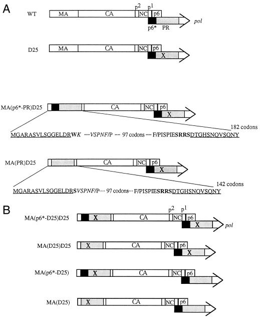
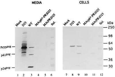
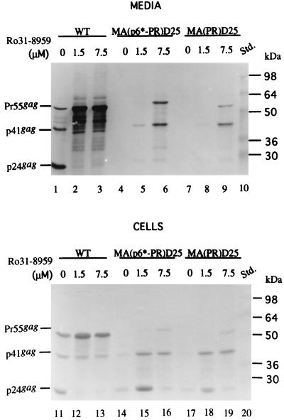
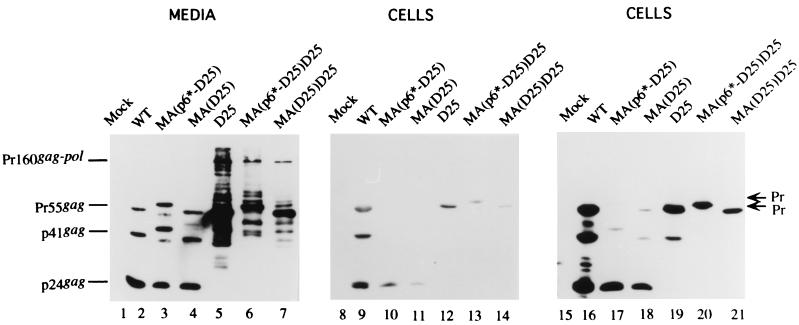
We constructed human immunodeficiency virus (HIV) mutants by replacing the matrix domain with sequences encoding the viral protease or p6* and protease. The chimeras retaining matrix myristylation and processing signals underwent efficient autoprocessing with severely defective particle budding. The budding defects of the chimeras were rescued by suppressing the chimera protease activity either through addition of an HIV protease inhibitor or through inactivating the chimera protease via a substitution mutation of the catalytic aspartic acid residue. This resulted in the release of chimeric virus-like particles with the density of a wild-type retrovirus particle. In addition, the assembly-competent but processing-defective chimeras produced proteolytically processed particles with significant reverse transcriptase activity when a downstream native pol gene was present. These results suggest that HIV has the potential to adapt heterologous sequences in place of the matrix sequence without major effects on virus-like particle budding. In addition, the positions of the protease and substrate accessibility may contribute significantly toward avoiding a premature Gag or Gag-Pol process, which leads to severe defects in both particle budding and incorporation.


Figures

FIG. 1
FIG. 1
Schematic presentation of the WT and mutant HIVgpt constructs. Mature WT processed Gag proteins and the p6∗ (black) and PR (stippled) domains of pol are indicated. The “X” indicates a PR-defective point mutation (D25→Asn). (A) The D25 mutant, which contains a substitution of an Asn residue for the PR catalytic Asp residue, is defective in Gag processing. The MA(p6∗-PR)D25 mutant contains a deletion of 105 codons and a replacement of the p6∗-PR coding sequence in the MA protein. The myristylation signal residues and a few residues in the C terminus of MA remain intact (underlined). Changed or added codons (boldfaced) and residues in the N and C termini of the PR domain are indicated. Upstream of PR, there are 45 codons (italics) of the p6∗ domain starting from the N-terminal 12th codon, K. The MA(PR)D25 mutant is identical to the MA(p6∗-PR)D25 mutant except that it contains only five C-terminal codons of p6∗. Instead of having 132 codons as in the WT MA protein, the MA(p6∗-PR) and the MA(PR) constructs contain a total of 182 and 142 codons in their MA regions, respectively. (B) Mutant constructs were derived from the constructs shown in panel A. MA(p6∗-D25)D25 and MA(D25)D25 were identical to MA(p6∗-PR)D25 and MA(PR)D25, respectively, except that the former two contain the PR-defective mutations (D25) in their chimera PR fragments. Recombination of the WT with MA(p6∗-D25)D25 and MA(D25)D25 yielded MA(p6∗-D25) and MA(D25), respectively.
FIG. 2
FIG. 2
Expression and processing of the chimeric proteins. 293T cells were transfected with the designated constructs. At 48 h posttransfection, cells and supernatants were collected for protein analysis. Supernatant samples (lanes 1 to 5) corresponding to 50% of the total samples and cell samples (lanes 7 to 11) corresponding to 5% of the total samples were fractionated by sodium dodecyl sulfate–10% polyacrylamide gel electrophoresis and electroblotted onto a nitrocellulose filter. HIV p24gag and p24gag-associated chimeric proteins were detected with mouse anti-p24gag monoclonal antibody at a 1:5,000 dilution, followed by a secondary alkaline phosphatase-conjugated sheep anti-mouse antibody at a 1:5,000 dilution, and alkaline phosphatase activity was determined. Positions of standard (Std.) molecular size markers (lanes 6 and 12) are indicated on the right, and those of HIV Gag proteins Pr55, p41, and p24 are shown on the left.
FIG. 3
FIG. 3
Release of the chimeras into the medium in the presence of an HIV PR inhibitor. 293T cells grown on 10-cm-diameter dish plates were transfected with the WT, MA(p6∗-PR)D25, and MA(PR)D25 HIVgpt constructs. At 18 h posttransfection, cells were split equally onto three 10-cm-diameter dishes and treated, respectively, with 0 μM (lanes 1, 4, 7, 11, 14, and 17), 1.5 μM (lanes 2, 5, 8, 12, 15, and 18), and 7.5 μM (lanes 3, 6, 9, 13, 16, and 19) concentrations of the HIV PR inhibitor Ro31-8959. Four hours later, the culture supernatants were removed and replaced with medium plus the designated concentration of the PR inhibitor. At 48 h after addition of the PR inhibitor, culture supernatants and cells were collected for protein analysis. Samples were fractionated by sodium dodecyl sulfate–10% polyacrylamide gel electrophoresis and subjected to immunoblot analysis with anti-p24gag antibody. Std., standards (lanes 10 and 20). Positions of the molecular size markers are indicated on the right, and those of the HIV Gag proteins Pr55, p41, and p24 are shown on the left.
FIG. 4
FIG. 4
Assembly and processing of chimeric particles. 293T cells were transfected with the designated plasmid. At 48 to 72 h posttransfection, supernatants and cells were prepared for Western immunoblotting. HIV-1 CA-associated proteins were detected by an enhanced-chemiluminescence detection system (Amersham). The primary antibody was an anti-p24gag monoclonal antibody used at a 1:5,000 dilution. The secondary antibody was a sheep anti-mouse horseradish peroxidase-conjugated antibody used at a 1:5,000 dilution. Positions of chimeric protein precursors are indicated on the right (arrows), and those of the HIV Gag proteins Pr55, p41, and p24 are shown on the left.

Similar articles

Cited by

References

    1. Chen Y-L, Ts'ai P-W, Yang C-C, Wang C-T. Generation of infectious virus particles by transient co-expression of human immunodeficiency virus type 1 gag mutants. J Gen Virol. 1997;78:2497–2501. - PubMed
    1. Freed E O. HIV Gag proteins: diverse functions in the virus life cycle. Virology. 1998;251:1–15. - PubMed
    1. Gelderblom H R. Assembly and morphology of HIV: potential effect of structure on viral function. AIDS. 1991;5:617–638. - PubMed
    1. Henderson L E, Bowers M A, Sowder II R C, Serabyn S A, Johnson D G, Bess J W, Jr, Arthur L O, Bryant D K, Fenselau C. Gag proteins of the highly replicative MN strain of human immunodeficiency virus type 1: posttranslational modifications, proteolytic processing, and complete amino acid sequences. J Virol. 1992;66:1856–1865. - PMC - PubMed
    1. Hunter E. Macromolecular interactions in the assembly of HIV and other retroviruses. Semin Virol. 1994;5:71–83.

Publication types