. 2000 Mar;105(5):567-75.
doi: 10.1172/JCI7497.

Hepatoma-derived growth factor stimulates smooth muscle cell growth and is expressed in vascular development

Affiliations

Hepatoma-derived growth factor stimulates smooth muscle cell growth and is expressed in vascular development

A D Everett et al. J Clin Invest. 2000 Mar.

Abstract

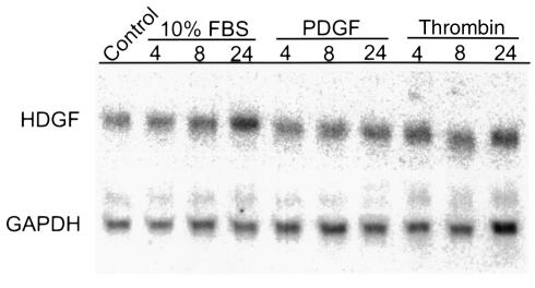
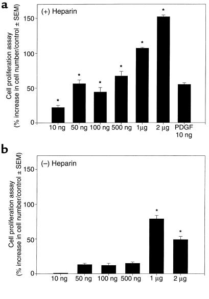
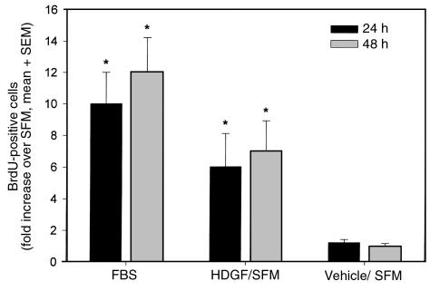
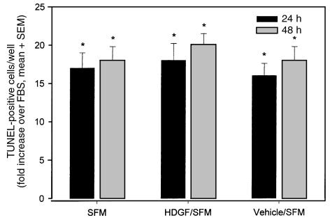
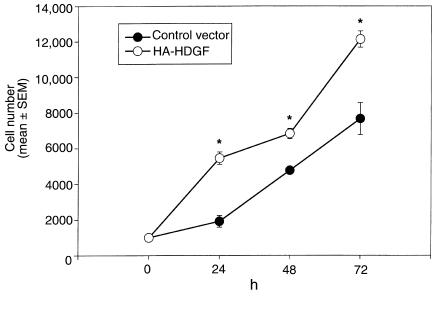
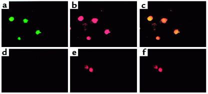
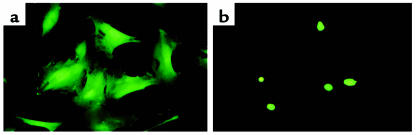
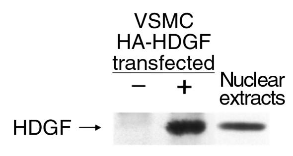
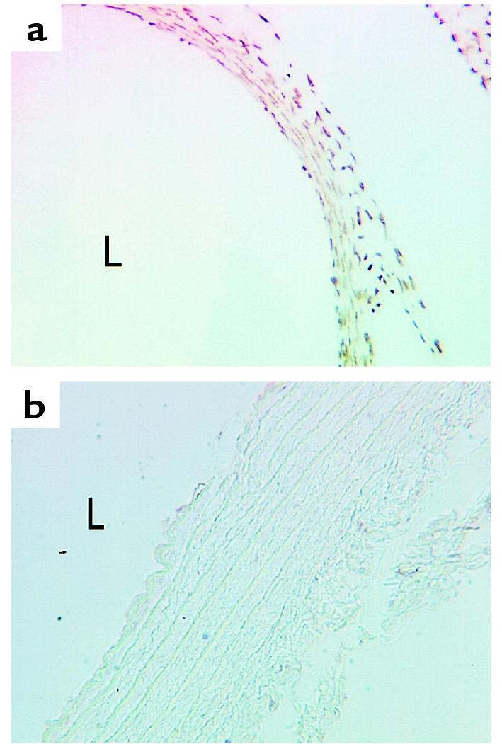
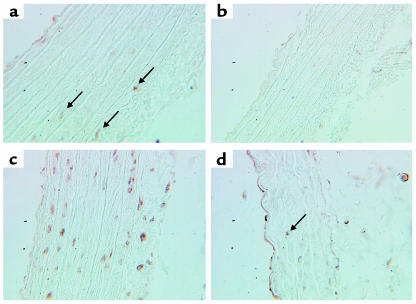
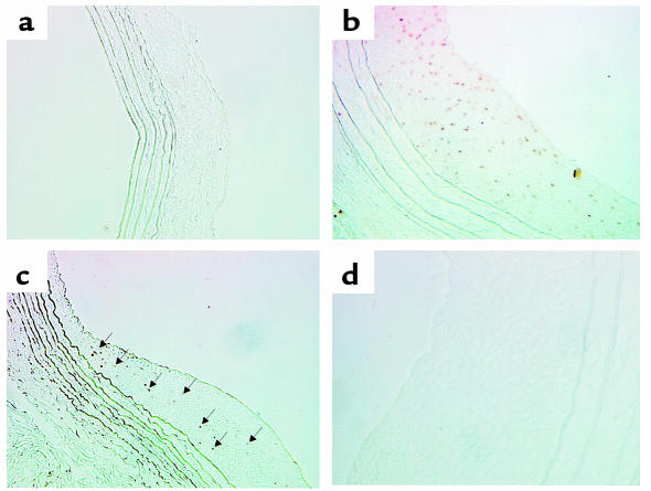
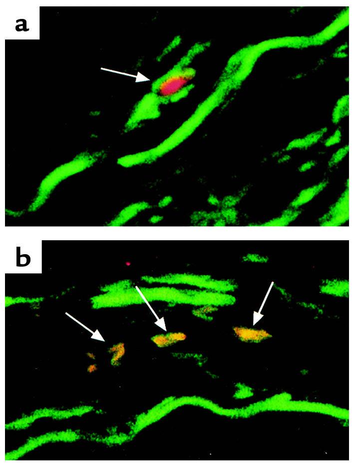
Hepatoma-derived growth factor (HDGF) is the first member identified of a new family of secreted heparin-binding growth factors highly expressed in the fetal aorta. The biologic role of HDGF in vascular growth is unknown. Here, we demonstrate that HDGF mRNA is expressed in smooth muscle cells (SMCs), most prominently in proliferating SMCs, 8-24 hours after serum stimulation. Exogenous HDGF and endogenous overexpression of HDGF stimulated a significant increase in SMC number and DNA synthesis. Rat aortic SMCs transfected with a hemagglutinin-epitope-tagged rat HDGF cDNA contain HA-HDGF in their nuclei during S-phase. We also detected native HDGF in nuclei of cultured SMCs, of SMCs and endothelial cells from 19-day fetal (but not in the adult) rat aorta, of SMCs proximal to abdominal aortic constriction in adult rats, and of SMCs in the neointima formed after endothelial denudation of the rat common carotid artery. Moreover, HDGF colocalizes with the proliferating cell nuclear antigen (PCNA) in SMCs in human atherosclerotic carotid arteries, suggesting that HDGF helps regulate SMC growth during development and in response to vascular injury.


Figures

Figure 1
Figure 1
Northern blot of HDGF mRNA expression in subconfluent vascular SMCs in response to serum, PDGF, or thrombin. SMCs were quiesced in SFM for 48 hours and treated with 10% FBS, PDGF (10 ng/mL), or thrombin (100 nM) for 4–24 hours. Total RNA (10 μg) was isolated and analyzed for HDGF mRNA expression, and GAPDH was used as a control for loading.
Figure 2
Figure 2
Effect of exogenous recombinant HDGF on SMC proliferation. SMCs in SFM with (a) or without (b) heparin were cultured for 72 hours with vehicle (control) or HDGF (10 ng–2 μg/mL) and assayed for cell number. Cell numbers per condition were expressed as percent increase over controls. For controls, n = 8; for HDGF, n = 4 per concentration. The entire experiment was repeated twice in 2 separate experiments. *P < 0.05 by one-way ANOVA control versus HDGF.
Figure 3
Figure 3
Effect of exogenous HDGF on BrdU uptake in SMCs. SMCs grown on coverslips in SFM were treated with SFM, 10% FBS, HDGF (100 ng/mL) or vehicle (PBS) for 24 or 48 hours and pulsed with BrdU for 6 hours. BrdU-positive nuclei were counted (n = 4 coverslips per group) and expressed as fold increase over SFM group as mean ± SEM. *P < 0.05 SFM versus FBS or HDGF by one-way ANOVA.
Figure 4
Figure 4
Effect of exogenous HDGF on the induction of apoptosis. SMC grown on coverslips were cultured in SFM alone, SFM + HDGF (100 ng/mL), SFM + vehicle (PBS), or 10% FBS for 24 or 48 hours. The TUNEL reaction was performed (n = 4 coverslips per group) and TUNEL-positive nuclei counted and expressed as the fold increase over the FBS group as mean ± SEM. *P < 0.05 versus the FBS group by one-way ANOVA.
Figure 5
Figure 5
Effect of endogenous overexpression of HDGF on SMC proliferation. SMCs were transfected with pKH3-HDGF and the transfection control plasmid pAdLox-GFP or the empty vector pKH3 and pAdLox-GFP. Forty-eight hours after transfection, cells were fluorescent cell sorted and replated at 1,000 cells per well. Cell proliferation was assayed by ELISA at 24, 48, and 72 hours after plating (n = 4 per group, repeated 3 times). Cell numbers are expressed as mean ± SEM. *P < 0.05 control versus HDGF-transfected groups.
Figure 6
Figure 6
Effect of HDGF overexpression in SMCs on BrdU uptake and cell localization. SMCs were transfected with HA-epitope–tagged HDGF (ac) or empty vector (df) and pulsed with BrdU as a marker of DNA synthesis for 12 hours. Cells were stained with an HA mAb and an FITC-conjugated secondary antibody to detect HDGF-transfected cells (green, a and d) and an alkaline phosphatase–conjugated anti-BrdU antibody to detect cells replicating their DNA (red, b and e). c is an overlay of a and b demonstrating colocalization (yellow) of HDGF to the nucleus of cells replicating their DNA. f is an overlay of d and e.
Figure 7
Figure 7
HDGF is targeted to the nucleus in living SMCs. SMCs were transfected with the empty vector (a, pK7-GFP) or (b) pK7-GFP-HDGF to express HDGF as an amino GFP fusion. HDGF expression was examined at 18 hours after transfection by fluorescent microscopy.
Figure 8
Figure 8
Native HDGF protein is located in the nucleus of cultured SMCs by Western analysis. Human adenocarcinoma cells were transfected with pKH3-HDGF (+) or empty vector alone (–). In lanes 1 and 2, recombinant HA-epitope–tagged HDGF was immunoprecipitated with an HA mAb from cellular extracts of transfected cells, and in lane 3 nuclear extracts from cultured SMCs were separated by 10% SDS-PAGE and blotted with an anti-HDGF polyclonal antibody.
Figure 9
Figure 9
HDGF expression in fetal and adult rat aorta. Nineteen-day fetal (a) and adult rat thoracic aorta (b) sections were immunostained for HDGF (1:1,000). HDGF-positive nuclei are brown with every nucleus positive in the fetal aorta (a), whereas only an occasional nucleus is seen to weakly stain in the adult aorta (b). L = lumen.
Figure 10
Figure 10
HDGF expression is increased in SMCs of the aorta after aortic constriction. HDGF immunostaining is shown in sham thoracic (a, arrows) and abdominal aorta (b) and in the thoracic aorta above the constriction (c, brown nuclei) and in the abdominal aorta below the constriction (d, arrow).
Figure 11
Figure 11
HDGF expression in the neointima after rat carotid artery balloon injury. HDGF immunostaining in sham (a) rat carotid arteries and at 14 days (b) and 28 days (c) after balloon injury. Brown nuclei (b and arrows in c) demonstrate positive HDGF immunostaining. d is a control serial section of b lacking the HDGF primary antibody.
Figure 12
Figure 12
HDGF expression in SMCs of atherosclerotic human carotid arteries. Representative sections from a human carotid surgical endarterectomy specimen were double immunostained for (a) HDGF (red) and α-smooth muscle actin (green) and (b) PCNA (green) and HDGF (red) to produce a yellow nucleus when the images are overlaid.

References

    1. Yoshida Y, Mitsumata M, Yamane T, Tomikawa M, Nishida K. Morphology and increased growth rate of atherosclerotic intimal smooth-muscle cells. Arch Pathol Lab Med. 1988;112:987–996. - PubMed
    1. Langlands K, Yin X, Anand G, Prochownik EV. Differential interactions of id proteins with basic-helix-loop-helix transcription factors. J Biol Chem. 1997;272:19785–19793. - PubMed
    1. Clowes AW, Reidy MA, Clowes MM. Kinetics of cellular proliferation after arterial injury. I. Smooth muscle growth in the absence of endothelium. Lab Invest. 1983;49:327–333. - PubMed
    1. Raines EW, Ross R. Smooth muscle cells and the pathogenesis of the lesions of atherosclerosis. Br Heart J. 1993;69(Suppl.):S30–S37. - PMC - PubMed
    1. McNamara CA, et al. Thrombin stimulates proliferation of cultured rat aortic smooth muscle cells by a proteolytically activated receptor. J Clin Invest. 1993;91:94–98. - PMC - PubMed

Publication types

MeSH terms