. 2000 Mar;105(5):597-605.
doi: 10.1172/JCI8047.

The ectodomain of the Notch3 receptor accumulates within the cerebrovasculature of CADASIL patients

Affiliations

The ectodomain of the Notch3 receptor accumulates within the cerebrovasculature of CADASIL patients

A Joutel et al. J Clin Invest. 2000 Mar.

Abstract

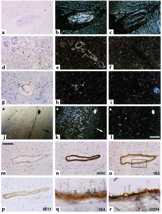
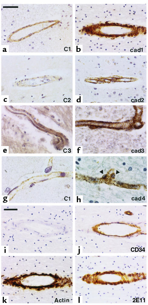
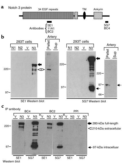
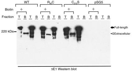
Mutations in Notch3 cause CADASIL (cerebral autosomal dominant adult onset arteriopathy), which leads to stroke and dementia in humans. CADASIL arteriopathy is characterized by major alterations of vascular smooth muscle cells and the presence of specific granular osmiophilic deposits. Patients carry highly stereotyped mutations that lead to an odd number of cysteine residues within EGF-like repeats of the Notch3 receptor extracellular domain. Such mutations may alter the processing or the trafficking of this receptor, or may favor its oligomerization. In this study, we examined the Notch3 expression pattern in normal tissues and investigated the consequences of mutations on Notch3 expression in transfected cells and CADASIL brains. In normal tissues, Notch3 expression is restricted to vascular smooth muscle cells. Notch3 undergoes a proteolytic cleavage leading to a 210-kDa extracellular fragment and a 97-kDa intracellular fragment. In CADASIL brains, we found evidence of a dramatic and selective accumulation of the 210-kDa Notch3 cleavage product. Notch3 accumulates at the cytoplasmic membrane of vascular smooth muscle cells, in close vicinity to but not within the granular osmiophilic material. These results strongly suggest that CADASIL mutations specifically impair the clearance of the Notch3 ectodomain, but not the cytosolic domain, from the cell surface.


Figures

Figure 1
Figure 1
Notch3 expression is restricted to vascular smooth muscle cells in human adult tissues. Notch3 expression was examined on paraffin sections from control individuals by in situ hybridization (al) and immunohistochemistry (mr). Sections of myocardium (ac), kidney (df), brain white matter (gi), and renal artery (jl) were hybridized with Notch3 antisense riboprobe (b, e, h, and k) and Notch3 sense riboprobe (c, f, i, and l). Kidney sections were hybridized with the HN3X riboprobes; all other sections with the SHB1 riboprobes. Bright fields (a, d, g, and j) and corresponding dark-field micrographs are shown. Notch3 is detected exclusively in the vessels (arrows) within the various parenchyma (ai), and primarily in the smooth muscle cell layer of the media or in vessels of the adventitia within the renal artery (jl). Small arrows in k indicate the internal elastica lamina, the thick arrow shows the external elastica lamina, and the star marks the luminal side. The bar in l represents 125 μm for ac, 100 μm for df, 50 μm for gi, and 200 μm for jl. (mr) Adjacent paraffin sections of brain frontal lobe were immunostained with primary antibodies recognizing the smooth muscle myosin heavy chain (mhc) (n), Notch3 extracellular epitopes 1E4 (o and q) and 2E11 (p), and the vascular endothelial cell marker CD34 (r). In m, primary antibody was omitted. Anti-Notch3 antibodies exclusively immunostained the smooth muscle cells (oq) but not the endothelial cells (arrowheads). Bar, 100 μm for mp and 20 μm for q and r (higher magnification of the boxed area of the vessel wall in o). Counterstaining was with toluidine blue (al) and with hematoxylin (mr).
Figure 2
Figure 2
Intense and granular appearance of Notch3 immunoreactivity in CADASIL brains. (ah) Paraffin sections from control (C1–C3; a, c, e, and g) and CADASIL brains (cad1–cad4; b, d, f, and h) including sections from white matter (a and b), cortex (c, d, g, and h), and brainstem (e and f), were probed with the 1E4 anti-Notch3 antibody. The examples shown are from 3 control individuals and 4 CADASIL brains matched for formalin fixation conditions and duration: 48 hours for control 1 (a and g) and CADASIL patient 1 (b); 0.5 months and 3 months for control 2 (c) and CADASIL patient 2 (d), respectively; 8 years for control 3 (e) and CADASIL patient 3 (f), and 1 year for CADASIL patient 4 (h). Notch3 immunoreactivity is much more intense within smooth muscle cells of vessels from CADASIL brains than in control brains (ah, right column compared with left column) for all durations of fixation. Granular and intense Notch3 immunostaining is also obvious within pericytes of capillaries (arrows, nuclei of pericytes) (h). Identical observations were obtained in brains from other donors (4 CADASIL patients, 2 controls). Bar, 50 μm for af and 20 μm for g and h. (il) Adjacent paraffin sections from CADASIL brain (patient 1) were probed with the 2E11 anti-Notch3 antibody (l), the anti-CD34 antibody (which is of the same isotype as the 1E4 and 2E11 antibodies) to identify the endothelial cells (j), the anti–smooth muscle α-actin antibody (to identify smooth muscle cells) (k), or without primary antibody (i). Bar, 50 μm. Counterstaining was with hematoxylin.
Figure 3
Figure 3
Proteolytic processing of wild-type Notch3. (a) Diagram of the Notch3 protein. Bars below the diagram indicate the various regions used as antigens to generate Notch3 antibodies (monoclonal antibodies 5E1, 11A1, and 5G7; polyclonal antibodies BC2 and BC4). TM, transmembrane domain. (b) Western blot analysis of transfected 293T cells and human control arterial tissue. Extracts were prepared from 293T cells transfected with human Notch1 (N1), Notch2 (N2), Notch3 (N3), or pSG5 vector (V). Fragment of a renal artery from a control individual was homogenized and then centrifuged at 16,000 g. The resulting pellet and supernatant were adjusted to 1× SDS-Laemmli buffer. Thirteen micrograms of transfected 293T cells and approximately 100 μg of pellet and supernatant (Sup) from the control artery were run on a 6% SDS-PAGE gel and incubated after transfer with 5E1 (left) and 5G7 (right). Positions of the 280-kDa full-length Notch3 (large black arrow) and the 210-kDa and 97-kDa processing products are indicated (open arrowhead and small arrow, respectively). Notice the weak expression level of Notch3 in arterial tissue, which required more than 1 hour of exposure as opposed to a few seconds for transfected cells. (c) Notch3 210-kDa and 97-kDa processing products are associated. Extracts from 293T cells transfected with Notch3 cDNA (N3) or vector alone (V) were immunoprecipitated with BC2 and BC4 polyclonal antibodies and with preimmune serum (PPI). Precipitated proteins were resolved by SDS-PAGE and immunoblotted with 5E1 and 5G7 antibodies. IP, immunoprecipitation. Migration of molecular-weight markers is shown to the left of each panel.
Figure 4
Figure 4
Selective accumulation of the 210-kDa cleavage product in CADASIL patients. (a and b) Western blot analysis of brain extracts from CADASIL and control individuals with antibodies raised against Notch3 extracellular domain. (a) Brain fragments were homogenized and centrifuged at 16,000 g; the resulting pellet and supernatant were adjusted to 1× SDS-Laemmli buffer. Approximately 100 μg of extracts were loaded per lane on a 6% SDS-PAGE gel, and were immunoblotted with the 5E1 antibody. Blots were stained with Ponceau S to confirm that equal amounts of proteins were loaded. Left panel, brain extracts (supernatant and pellet) from CADASIL patient 1 and control individuals 2 and 5. Middle panel, brain extract (pellet) from CADASIL patient 1 and extract from 293T cells transfected with Notch3 cDNA (N3). Right panel, brain extracts (pellet) from CADASIL patients 1, 8, and 9. Position of the 210-kDa Notch3 protein (open arrowhead) detected in CADASIL brain extracts is indicated. The asterisk marks a band of unknown significance (cross-reacting material or, most likely, post-lysis degradation product). (b) Brain extracts from CADASIL patient 1 (pellet) and control individual 2 (pellet), and extracts from 293T cells transfected with Notch3 cDNA (N3) were run on a 6% SDS-PAGE gel and immunoblotted with the 11A1 antibody (left panel) or without primary antibody (–) (right panel). (c) The 97-kDa Notch3 protein is not detected in either CADASIL or control brains. Whole lysates were prepared from brains of CADASIL patient 1 and control individual 5, and from a renal artery of a control subject. Approximately 100 μg of each extract was loaded on a 6% SDS-PAGE gel and immunoblotted with the 5G7 antibody (bottom) and the 5E1 antibody (top). Positions of the 210-kDa and the 97-kDa processing products are indicated (open arrowhead and small arrow, respectively). (d) Western blot analysis of extracts from CADASIL and control arterial tissues. Whole lysates were prepared from mesenteric arteries of CADASIL patient 1 and a renal artery of a control subject. Extracts were resolved on a 6% SDS-PAGE gel and immunoblotted with the 5G7 antibody (right) and the 5E1 antibody (left). The asterisk marks a band of unknown significance (cross-reacting material or, most likely, a post-lysis degradation product). Whole lysates were used in experiments shown in c and d to avoid missing any soluble Notch3 intracellular fragment. (e) Western blot analysis of extracts from CADASIL and control brains and from control arterial tissue under reducing and nonreducing conditions. Extracts from CADASIL and control brains and from a control artery were solubilized in SDS-Laemmli buffer with or without βME and were resolved on a 6% SDS-PAGE gel. Both stacking (bracket) and separating gels were immunoblotted with the 5E1 antibody. Under reducing conditions (βME+), the 210-kDa Notch3 protein is detected in both control artery and CADASIL brain (open arrowhead). Under nonreducing conditions (βME–), no band is detected in the CADASIL brain; a protein of approximately 280 kDa (large open arrow) is detected in the control artery. This protein probably corresponds to the associated extracellular and intracellular domains. Cross-reacting material is caught at the transition between the stacking and separating gels.
Figure 5
Figure 5
Wild-type and mutant Notch3 proteins expressed at the cell surface of transfected 293T cells are cleaved. 293T cells transfected with wild-type Notch3 (WT), mutant Notch3 (R90C and C212S), and vector alone (pSG5) were incubated with sulfo-NHS-biotin (+) or were mock treated. Cells were lysed in RIPA buffer, and extracts were loaded on a 6% SDS-PAGE gel either directly (T fraction, 15% of the extract) or after incubation on streptavidin-agarose beads (B fraction, 85% of the extract). Extracts were then immunoblotted with the 5E1 antibody. Positions of the 280-kDa full-length Notch3 extracellular domain (black arrow) and the 210-kDa Notch3 extracellular domain (arrowhead) are indicated.
Figure 6
Figure 6
Notch3 immunogold labeling is highly clustered at the cell surface of smooth muscle cells. (a) Vessel within the frontal lobe of CADASIL patient 1, processed for electron microscopy using standard procedure. Outline of a smooth muscle cell (m) is indicated by open small arrows. Granular osmiophilic deposits (large arrows) are observed within the basal lamina (square), in close contact with the cytoplasmic membrane of this smooth muscle cell. ×13,000. (b) Vessel within the occipital lobe from CADASIL patient 1, processed for immunoelectron microscopy with the 1E4 antibody. Numerous granular osmiophilic deposits (large arrows) are observed within the basal lamina (square). These are nested within invaginations of the smooth muscle cells (m), or are spread far away within the vessel wall (stars). Notch3 immunolabeling, which appears as black dots due to silver enhancement, is highly clustered at the cell membrane of smooth muscle cells (open small arrows) closely arranged around the deposits, which are not labeled. ×20,000. Ultrathin sections stained with uranyl acetate and lead citrate. ec, endothelial cell; Lu, lumen.

Comment in

References

    1. Joutel A, et al. Notch3 mutations in CADASIL, an hereditary adult-onset condition causing stroke and dementia. Nature. 1996;383:707–710. - PubMed
    1. Tournier-Lasserve E, Iba-Zizen MT, Romero N, Bousser MG. Autosomal dominant syndrome with stroke-like episodes and leukoencephalopathy. Stroke. 1991;22:1297–1302. - PubMed
    1. Tournier-Lasserve E, et al. Cerebral autosomal dominant arteriopathy with subcortical infarcts and leukoencephalopathy maps to chromosome 19q12. Nat Genet. 1993;3:256–259. - PubMed
    1. Chabriat H, et al. Clinical spectrum of CADASIL: a study of 7 families. Lancet. 1995;346:934–939. - PubMed
    1. Baudrimont M, Dubas F, Joutel A, Tournier-Lasserve E, Bousser MG. Autosomal dominant leukoencephalopathy and subcortical ischemic stroke. A clinicopathological study. Stroke. 1993;24:122–125. - PubMed

Publication types

MeSH terms