. 2000 Mar;105(5):625-31.
doi: 10.1172/JCI8748.

Use of autoantigen-knockout mice in developing an active autoimmune disease model for pemphigus

Affiliations

Use of autoantigen-knockout mice in developing an active autoimmune disease model for pemphigus

M Amagai et al. J Clin Invest. 2000 Mar.

Abstract

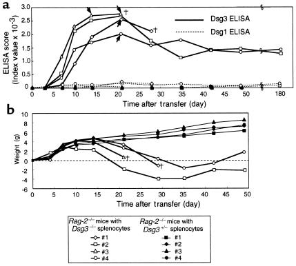
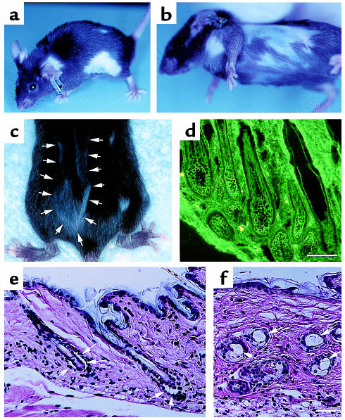
The development of experimental models of active autoimmune diseases can be difficult due to tolerance of autoantigens, but knockout mice, which fail to acquire tolerance to the defective gene product, provide a useful tool for this purpose. Using knockout mice lacking desmoglein 3 (Dsg3), the target antigen of pemphigus vulgaris (PV), we have generated an active disease model for this autoantibody-mediated disease. Dsg3(-/-) mice, but not Dsg3(+/-) littermates, produced anti-Dsg3 IgG that binds native Dsg3, when immunized with recombinant mouse Dsg3. Splenocytes from the immunized Dsg3(-/-) mice were then adoptively transferred into Rag-2(-/-) immunodeficient mice expressing Dsg3. Anti-Dsg3 IgG was stably produced in the recipient mice for more than 6 months without further boosting. This IgG bound to Dsg3 in vivo and disrupted the cell-cell adhesion of keratinocytes. Consequently, the recipient mice developed erosions in their oral mucous membranes with typical histologic findings of PV. In addition, the recipient mice showed telogen hair loss, as found in Dsg3(-/-) mice. Collectively, the recipient mice developed the phenotype of PV due to the pathogenic anti-Dsg3 IgG. This model will be valuable for developing novel therapeutic strategies. Furthermore, our approach can be applied broadly for the development of various autoimmune disease models.


Figures

Figure 1
Figure 1
Anti-Dsg3 IgG antibodies that can bind the native Dsg3 in vivo are produced in Dsg3–/– mice, but not in Dsg3+/– mice. (a) Dsg3–/– mice and their Dsg3+/– littermates were immunized with mouse rDsg3, and the ELISA titers against rDsg3 were measured over time. Mice were primed by intraperitoneal injection of purified mouse rDsg3 in complete Freund’s adjuvant on day 0 (arrow). They were subsequently boosted with mouse rDsg3 in incomplete Freund’s adjuvant (solid triangles), and then injected with the mouse rDsg3 without adjuvant (open triangles). (b) A mouse keratinocyte cell line, PAM212, was incubated with mouse serum samples in culture media in a CO2 incubator for 30 minutes. After being washed and fixed with methanol, bound mouse IgG was revealed with FITC-conjugated goat anti-mouse IgG antibodies. Sera from immunized Dsg3–/– mice (left), but not from their Dsg3+/– littermates (right), stained the cell-cell contact sites of cultured keratinocytes. Bar, 50 μm.
Figure 2
Figure 2
Stable production of anti-Dsg3 IgG in mice given immunized Dsg3–/– splenocytes. Splenocytes were isolated from immunized Dsg3–/– or Dsg3+/– mice and then transferred to Rag-2–/– mice by intravenous injection into the tail vein. (a) Circulating anti-Dsg3 IgG was detected in the mice that received Dsg3–/– splenocytes (open symbols, solid line), but not in the recipients of Dsg3+/– splenocytes (solid symbols). The antibody production persisted for more than 6 months. There was no apparent reactivity against Dsg1 (dashed line). Arrows indicate the day when the hair loss phenotype became apparent. (b) Weight change in the recipient Rag-2–/– mice is shown by plotting weight against time.†indicates the death of a mouse.
Figure 3
Figure 3
Rag-2–/– mice injected with immunized Dsg3–/– splenocytes develop the PV phenotype. (a) Mice receiving Dsg3–/– splenocytes (bottom) were significantly smaller than mice given Dsg3+/– splenocytes (top) 25–35 days after the adoptive transfer. (b) Some mice injected with Dsg3–/– splenocytes developed crusted erosions around the snout and cheeks, where mice normally scratch. (cj) Histologic and immunopathologic examination of Rag-2–/– recipient mice and patients with PV. In vivo IgG deposition on keratinocyte cell surfaces was observed in the skin (c, the skin around the snout) and mucous membranes (d, hard palate) of Rag-2–/– mice given Dsg3–/– splenocytes, just as is found in patients with PV (f, esophagus biopsy specimen). In contrast, there was no in vivo IgG deposition in the mice given Dsg3+/– splenocytes (e, hard palate). The mice that received Dsg3–/– splenocytes developed intraepithelial blisters just above the basal layers in mucosal epithelium (g, hard palate; h, upper esophagus), which is a typical histologic finding in PV patients (j, skin). There was no apparent sign of acantholysis in the mice with Dsg3+/– splenocytes (i, upper esophagus). Bars = 50 μm.
Figure 4
Figure 4
Hair loss phenotype of Rag-2–/– mice given splenocytes from immunized Dsg3–/– mice. (a, b) The Rag-2–/– mice given Dsg3–/– splenocytes showed patchy hair loss, which first became apparent around day 15–25. (c) New hair growing in a bald area in a patchy pattern (arrows). (d) Intense in vivo IgG deposition was noticed on the cell surface of keratinocytes surrounding the telogen hair club. (e, f) Cleft formation between the cells surrounding the telogen club and the basal layer of the outer root sheath epithelium (e, arrows) and empty, dilated telogen hair follicles (f, arrows). Bars = 100 μm.

References

    1. MacDonald HR. Mechanisms of immunological tolerance. Science. 1989;246:982. - PubMed
    1. Goodnow CC. Balancing immunity and tolerance: deletion and tuning lymphocyte repertoires. Proc Natl Acad Sci USA. 1996;93:2264–2271. - PMC - PubMed
    1. Bach JF, Koutouzov S, van Endert PM. Are there unique autoantigens triggering autoimmune diseases? Immunol Rev. 1998;164:139–155. - PubMed
    1. Stanley, J.R. 1998. Pemphigus. In Dermatology in general medicine. I.M. Freedberg et al., editors. McGraw-Hill. New York, NY. 654–666.
    1. Amagai M, Klaus-Kovtun V, Stanley JR. Autoantibodies against a novel epithelial cadherin in pemphigus vulgaris, a disease of cell adhesion. Cell. 1991;67:869–877. - PubMed

Publication types

MeSH terms

Associated data